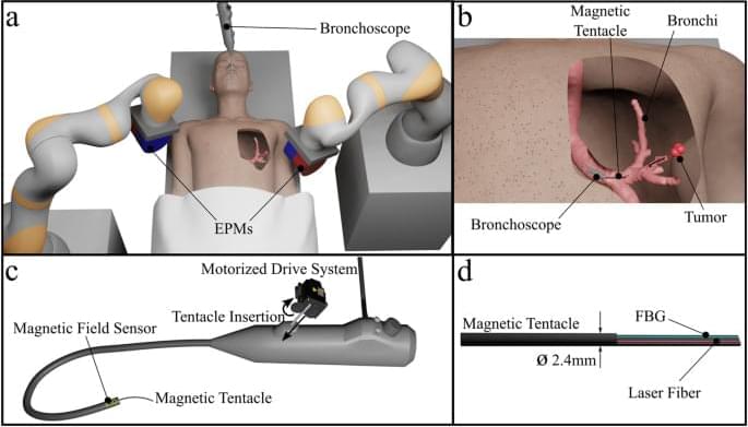
All navigations reported in Fig. 2 were performed autonomously within 150 s and without intraoperative imaging. Specifically, each navigation was performed according to the pre-determined optimal actuation fields and supervised in real time by intraoperative localization. Therefore, the set of complex navigations performed by the magnetic tentacle was possible without the need for exposure to radiation-based imaging. In all cases, the soft magnetic tentacle is shown to conform by design to the anatomy thanks to its low stiffness, optimal magnetization profile and full-shape control. Compared to a stiff catheter, the non-disruptive navigation achieved by the magnetic tentacle can improve the reliability of registration with pre-operative imaging to enhance both navigation and targeting. Moreover, compared to using multiple catheters with different pre-bent tips, the optimization approach used for the magnetic tentacle design determines a single magnetization profile specific to the patient’s anatomy that can navigate the full range of possible pathways illustrated in Fig. 2. Supplementary Movies S1 and S2 report all the experiments. Supplementary Movie S1 shows the online tracking capabilities of the proposed platform.
In Table 1, we report the results of the localization for four different scenarios. These cases highlight diverse navigations in the left and right bronchi. The error is referred to as the percentage of tentacles outside the anatomy. This was computed by intersecting the shape of the catheter, as predicted by the FBG sensor, and the anatomical mesh grid extracted from the CT scan. The portion of the tentacle within the anatomy was measured by using “inpolyhedron” function in MATLAB. In Supplementary Movie S1, this is highlighted in blue, while the section of the tentacle outside the anatomy is marked in red. The error in Table 1 was computed using the equation.


Comments are closed.