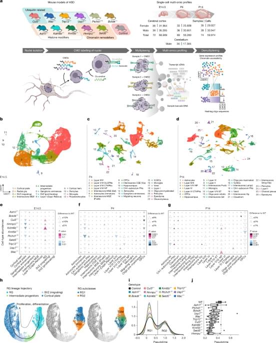
Using single-nucleus multi-omic sequencing, diverse autism spectrum disorder-linked gene mutations converge on transient, stage-specific disruptions in early brain development, and highlight sex-specific gene expression alterations.
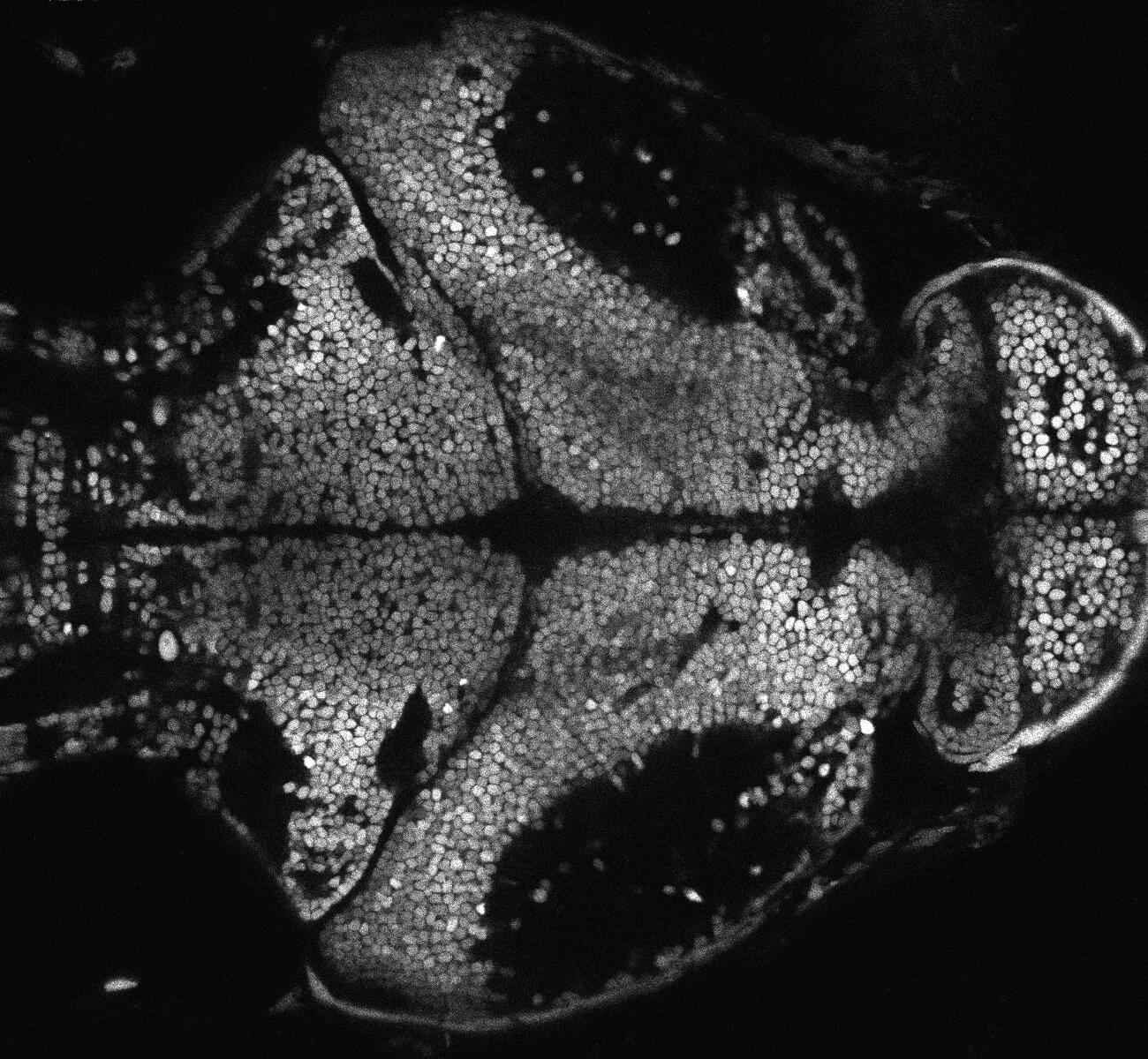
The altered presence of tiny fragments of neuronal genes, called microexons, causes hyperarousal in zebrafish. This is the main conclusion of an international study led by Pompeu Fabra University (UPF) and the Center for Genomic Regulation (CRG). An abnormal pattern of neural microexon presence leads to a hyperarousal state characterized by heightened neural activity and insomnia, commonly associated with stress but also with neurodevelopmental disorders.
Arousal regulation is highly conserved in evolution. Therefore, this finding could help researchers understand the mechanism underlying some human neurodevelopmental disorders, such as autism and schizophrenia, conditions associated with microexon mutations.
To survive, animals need to be ready to react to external and internal stimuli. This activation of the central nervous system, arousal, is highly conserved throughout the animal kingdom.
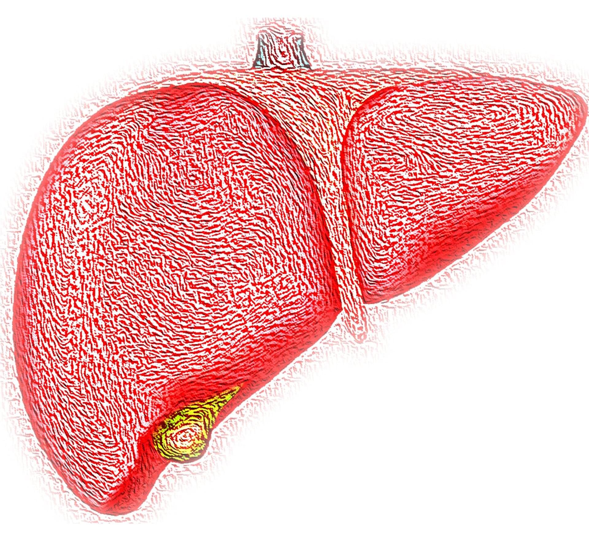
A new gene therapy has been used to successfully treat a deadly childhood liver disease in mice that model the disease, according to researchers at UCL and Great Ormond Street Hospital. Arthrogryposis, renal dysfunction and cholestasis (ARC) syndrome is a lethal genetic disorder usually caused by a lack of the VPS33B protein, with children diagnosed with the condition rarely living beyond their first year of life.
Now, in a study published in Nature Communications, the UCL-GOSH team found that by injecting a healthy version of the gene into the body, they can treat the condition in mice lacking VPS33B. Crucially, the final version of the treatment, which specifically targeted the liver cells, caused no harm. In the earlier versions, the genes became abnormally activated and caused cancerous cells to grow and expand in some cases.
While more tests must be done before the treatment can be tested in humans, the researchers’ breakthrough offers hope to babies with this devastating disorder and their families. In the UK, as many as six pregnancies per year might be affected by ARC syndrome. Furthermore, the findings may promote improved understanding of why some treatments may cause cancer.
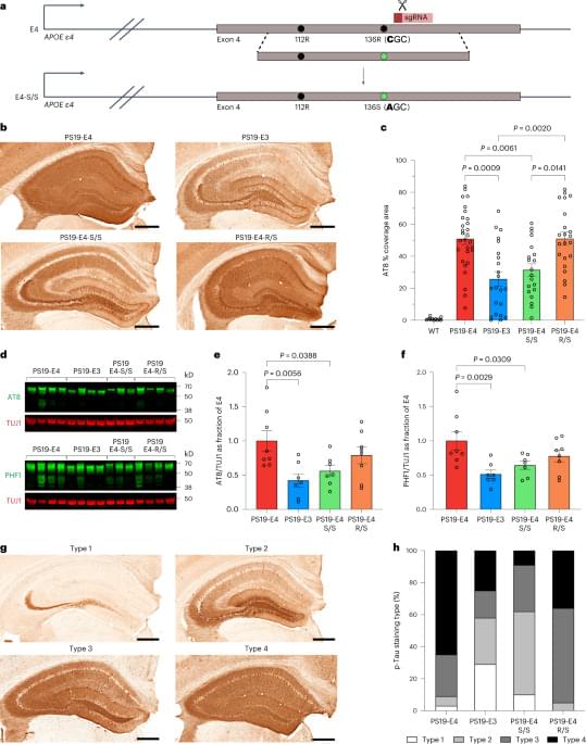
Nelson et al. present a detailed biomolecular study of how the APOE-R136S mutation protects against Alzheimer’s disease (AD) in mice and in patient-derived cells. Lots of data on glial contributions and transcriptomic changes. I see this as an excellent target for gene therapies aiming to combat AD. So do the folks at Lexeo Therapeutics (an exciting company you should check out!)
Nelson et al. report that the APOE-R136S mutation protects against APOE4-promoted Alzheimer’s disease pathologies, including phosphorylated Tau accumulation, neuroinflammation and neurodegeneration, in mouse and human neuron models.

Aging is a universal biological process, yet the reasons why some individuals live significantly longer and healthier lives have long puzzled scientists. Among the genes linked to exceptional longevity, FOXO3 consistently stands out as one of the most influential “master controllers” of cellular resilience. This single transcription factor integrates signals from stress, metabolism, DNA repair, and stem cell biology, orchestrating a vast genetic program that determines how cells survive, adapt, or age [1].
In recent years, interest in FOXO3 has surged across aging research, regenerative medicine, oncology, and precision therapeutics. Variants of the FOXO3 gene are strongly associated with centenarian populations worldwide, while disruptions in its regulatory network contribute to multiple disorders, including cancer, neurodegeneration, metabolic decline, and tissue degeneration. With advances in computational biology and pathway analysis, it is now possible to map FOXO3’s complex signaling network and uncover new therapeutic strategies.
This blog post explores FOXO3’s multifaceted biological roles, its influence on disease, and what our curated data from TRANSFAC®, TRANSPATH®, and HumanPSD™ reveals about the FOXO3 regulatory network. The goal is to provide a scientifically rich yet accessible overview that sparks curiosity among researchers studying aging, longevity, and systems-level biology.
What if a single sentence could carry two completely different meanings, one when read forward and another when read backward? In a new study, researchers at Arizona State University have discovered a biological version of this idea. Working with the mitochondria of a tiny insect called the citrus mealybug, the team found that the same stretch of DNA can carry two different genes—sets of genetic instructions used by the cell—with one encoded on each strand of the DNA’s ladder-like structure.
The finding expands scientists’ understanding of how DNA can store genetic information and helps solve a mystery that has puzzled researchers for years. The findings are published in the journal Proceedings of the National Academy of Sciences.
“This kind of paper is what makes running a lab so fun. Born from a spark of individual brilliance—not mine—but accomplished as a collective effort,” says John McCutcheon. “The idea that these two critically important genes could be mirrored on the same piece of DNA has been around a long time, and so it’s a thrill to be part of the team that proved this speculative idea was, in fact, reality.”
Brody is professor of neuroscience and molecular biology at Princeton University and a Howard Hughes Medical Institute Investigator. His research focuses is on novel quantitative behaviors that allow exploring high-level cognitive questions using powerful emerging tools for studying neural mechanisms in rodents. Brody’s group uses rats to investigate the neural bases of decision making, working memory, and executive control, using a combination of high-throughput semiautomated behavior as well as computational, electrophysiological, pharmacological and optogenetic methods.
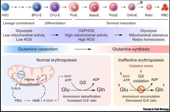
Red blood cell (RBC) production, or erythropoiesis, serves as a paradigm for studying cellular differentiation in both physiological and pathological contexts. While the transcriptional and epigenetic programs controlling erythropoiesis are well characterized, the metabolic regulation of this complex process remains underexplored. Recent discoveries that pyruvate kinase activators improve outcomes in sickle cell disease and thalassemia underscore the therapeutic potential of targeting metabolism in RBC disorders. However, further progress is limited by an incomplete understanding of the metabolic networks supporting erythropoiesis and RBC function.
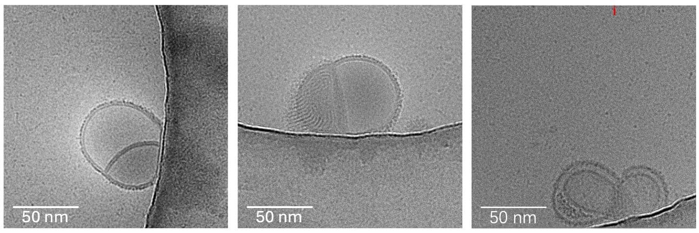
Researchers at the University of Houston’s College of Pharmacy have discovered an unexpectedly simple strategy to improve the performance of mRNA vaccines and gene therapeutics: adding salt. The findings, published in Small, address one of the biggest challenges facing modern gene medicine—getting fragile therapeutic material to the right place inside cells.
“We are introducing salt-loaded lipid nanoparticles as a novel and broadly applicable design principle for gene delivery,” said Fanfei Meng, assistant professor and Presidential Frontier Faculty member in the Department of Pharmacological and Pharmaceutical Sciences. “What makes this exciting is that we can significantly improve delivery efficiency without needing to invent entirely new materials.”
Lipid nanoparticles, or LNPs, are tiny fat-based delivery vehicles widely used to transport fragile genetic material into cells. They became widely recognized during the COVID-19 pandemic through mRNA vaccines developed by Moderna and Pfizer. Today, scientists are also using LNPs to develop new treatments for cancer, rare diseases and genetic disorders.