The technique, which used genetically healthy donor cells, prolonged life and function in mice with a disease similar to Tay-Sachs. It may help with other neurodegenerative diseases like Alzheimer’s.
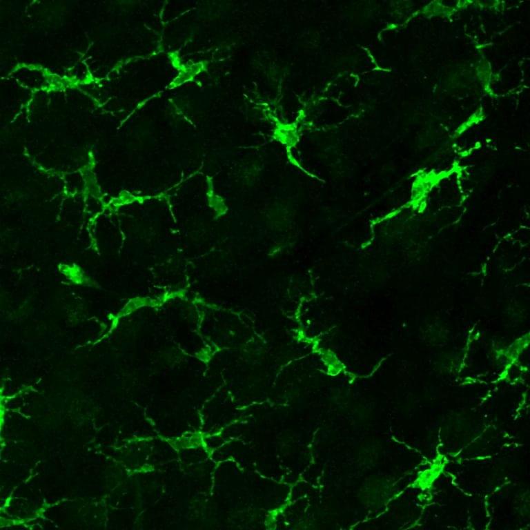

The technique, which used genetically healthy donor cells, prolonged life and function in mice with a disease similar to Tay-Sachs. It may help with other neurodegenerative diseases like Alzheimer’s.
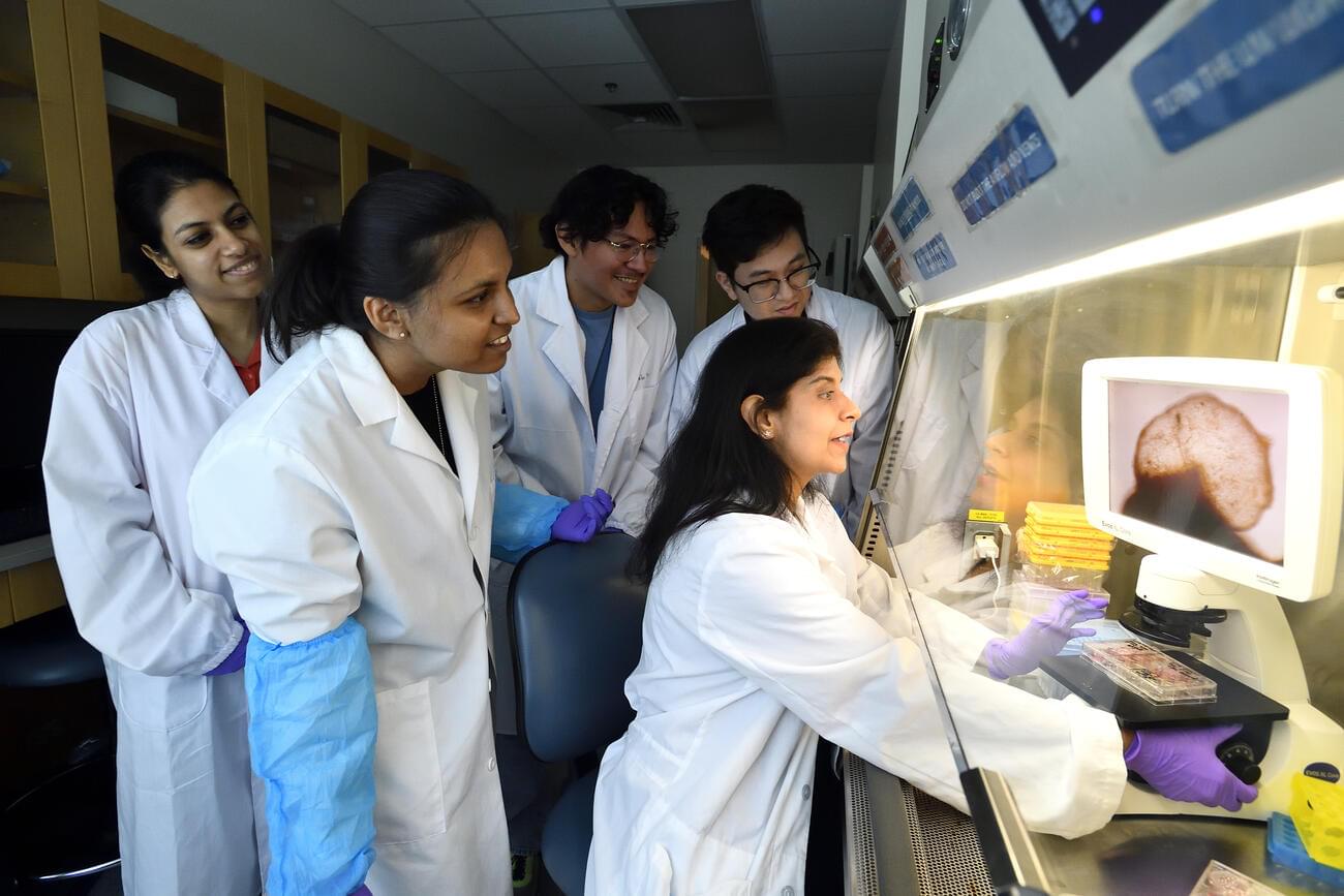
Johns Hopkins University researchers have grown a novel whole-brain organoid, complete with neural tissues and rudimentary blood vessels—an advance that could usher in a new era of research into neuropsychiatric disorders such as autism.
“We’ve made the next generation of brain organoids,” said lead author Annie Kathuria, an assistant professor in JHU’s Department of Biomedical Engineering who studies brain development and neuropsychiatric disorders. “Most brain organoids that you see in papers are one brain region, like the cortex or the hindbrain or midbrain. We’ve grown a rudimentary whole-brain organoid; we call it the multi-region brain organoid (MRBO).”
The venerable Ralph Merkle joins Max Marty on stage at Vitalist Bay.
Key topics covered:
- Why cryonics is still “0.00001%” of people despite decades of advocacy — and Merkle’s admission that believing rational arguments would work was his biggest mistake.
- The wild Dora Kent story.
- The organizational split in 1992 that affected growth for over a decade.
- Why Merkle’s success probability hasn’t changed since the 80s.
- Whether preserving your information pattern is enough or if you need “continuity of consciousness”.
And more.
Links:
• Cryosphere Discord server: https://discord.com/invite/ndshSfQwqz.
• Cryonics subreddit: https://www.reddit.com/r/cryonics/
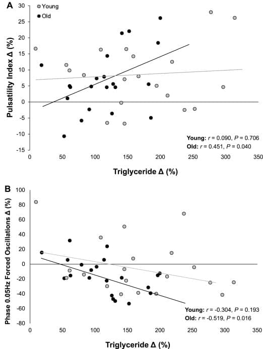
Dietary fat is an important part of our diet. It provides us with a concentrated source of energy, transports vitamins and when stored in the body, protects our organs and helps keep us warm. The two main types of fat that we consume are saturated and unsaturated (monounsaturated and polyunsaturated), which are differentiated by their chemical composition.
But these fats have different effects on our body. For example, it is well established that eating a meal that is high in saturated fat, such as that self-indulgent Friday night takeaway pizza, can be bad for our blood vessels and heart health. And these effects are not simply confined to the heart.
The brain has limited energy stores, which means it is heavily reliant on a continuous supply of blood delivering oxygen and glucose to maintain normal function.
One of the ways the body maintains this supply is through a process known as “dynamic cerebral autoregulation”. This process ensures that blood flow to the brain remains stable despite everyday changes in blood pressure, such as standing up and exercising. It’s like having shock absorbers that help keep our brains cool under pressure.
But when this process is impaired, those swings in blood pressure become harder to manage. That can mean brief episodes of too little or too much blood reaching the brain. Over time, this increases the risk of developing conditions like stroke and dementia.
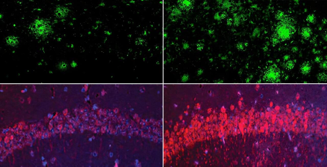
What is the earliest spark that ignites the memory-robbing march of Alzheimer’s disease? Why do some people with Alzheimer’s-like changes in the brain never go on to develop dementia? These questions have bedeviled neuroscientists for decades.
Now, a team of researchers at Harvard Medical School may have found an answer: lithium deficiency in the brain.
The work, published in Nature, shows for the first time that lithium occurs naturally in the brain, shields it from neurodegeneration, and maintains the normal function of all major brain cell types.

In a major new finding almost a decade in the making, researchers at Harvard Medical School say they’ve found a key that may unlock many of the mysteries of Alzheimer’s disease and brain aging — the humble metal lithium.
Lithium is best known to medicine as a mood stabilizer given to people who have bipolar disorder and depression. It was approved by the US Food and Drug Administration in 1970, but it was used by doctors to treat mood disorders for nearly a century beforehand.
Now, for the first time, researchers have shown that lithium is naturally present in the body in tiny amounts and that cells require it to function normally — much like vitamin C or iron. It also appears to play a critical role in maintaining brain health.
Researchers from the Broad Institute and Mass General Brigham have shown that a low-oxygen environment—similar to the thin air found at Mount Everest base camp—can protect the brain and restore movement in mice with Parkinson’s-like disease.
The new research, in Nature Neuroscience, suggests that cellular dysfunction in Parkinson’s leads to the accumulation of excess oxygen molecules in the brain, which then fuel neurodegeneration—and that reducing oxygen intake could help prevent or even reverse Parkinson’s symptoms.
“The fact that we actually saw some reversal of neurological damage is really exciting,” said co-senior author Vamsi Mootha, an institute member at the Broad, professor of systems biology and medicine at Harvard Medical School, and a Howard Hughes Medical Institute investigator in the Department of Molecular Biology at Massachusetts General Hospital (MGH), a founding member of the Mass General Brigham healthcare system.

Metaphors are a fundamental aspect of human language and cognition, allowing us to understand complex concepts and relationships by mapping them onto more familiar and concrete domains. However, the nature of metaphors and how they work is still not well understood.
In a new paper published in PLOS Complex Systems, Max-Planck-Institute for Mathematics in the Sciences researchers Marie Teich and Wilmer Leal together with director Jürgen Jost have developed a formal framework and large-scale empirical methodology to analyze metaphors and their role in conceptual metaphor theory.
The study confirms the fundamental assumption in conceptual metaphor theory that metaphors are enduring linguistic and cognitive structures, not merely rhetorical figures. Using complex systems tools, the researchers identified a metaphor network with distinctions between abstract and concrete categories, and two significant metaphorical processes: mappings from concrete to abstract topics and the emergence of new mappings between concrete domains.
