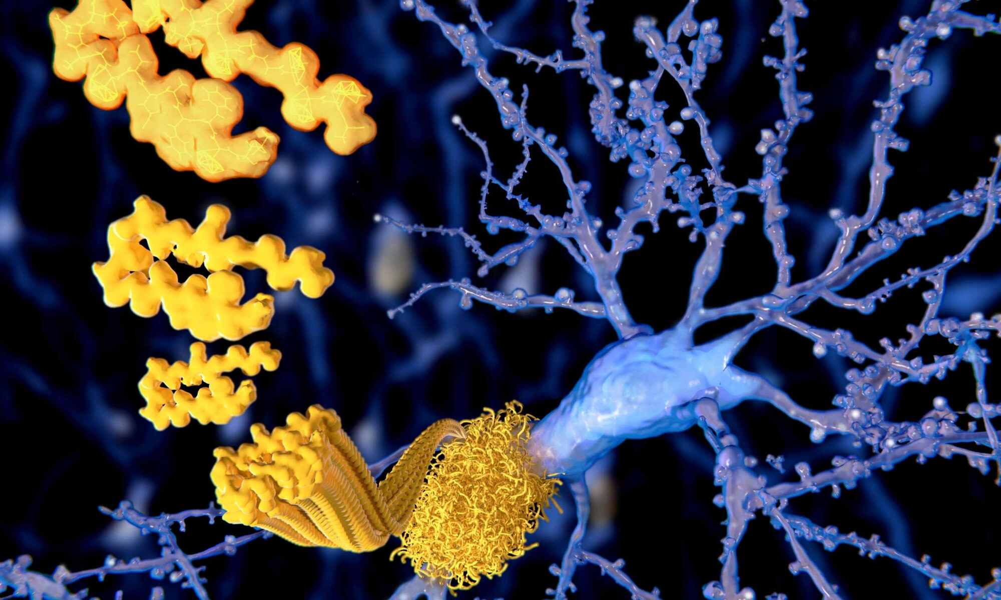
Motherhood appears to lead to long-lasting increases in gray matter density in the brain, particularly in cognitive and visual areas, which may protect against aging.


#theoryofeverything #consciousness #philosophy Hey Every0ne, happy new year! Tonight we will discuss a talk on Theories of Everything @TheoriesofE…

Humans tend to put our own intelligence on a pedestal. Our brains can do math, employ logic, explore abstractions, and think critically. But we can’t claim a monopoly on thought. Among a variety of nonhuman species known to display intelligent behavior, birds have been shown time and again to have advanced cognitive abilities. Ravens plan for the future, crows count and use tools, cockatoos open and pillage booby-trapped garbage cans, and chickadees keep track of tens of thousands of seeds cached across a landscape. Notably, birds achieve such feats with brains that look completely different from ours: They’re smaller and lack the highly organized structures that scientists associate with mammalian intelligence.
“A bird with a 10-gram brain is doing pretty much the same as a chimp with a 400-gram brain,” said Onur Güntürkün, who studies brain structures at Ruhr University Bochum in Germany. “How is it possible?”
Researchers have long debated about the relationship between avian and mammalian intelligences. One possibility is that intelligence in vertebrates—animals with backbones, including mammals and birds—evolved once. In that case, both groups would have inherited the complex neural pathways that support cognition from a common ancestor: a lizardlike creature that lived 320 million years ago, when Earth’s continents were squished into one landmass. The other possibility is that the kinds of neural circuits that support vertebrate intelligence evolved independently in birds and mammals.
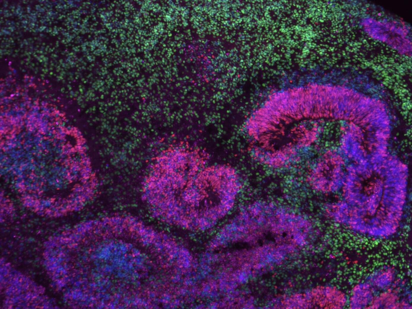
The human brain is known to contain a wide range of cell types, which have different roles and functions. The processes via which cells in the brain, particularly its outermost layer (i.e., the cerebral cortex), gradually become specialized and take on specific roles have been the focus of many past neuroscience studies.
Researchers at the University of California Los Angeles (UCLA) analyzed different datasets collected using single-cell transcriptomics, a technique to study gene expression in individual cells, to map the emergence of different cell types during the brain’s development.
Their findings, published in Nature Neuroscience, unveil gene “programs” that drive the specialization of cells in the human cerebral cortex.


“I think, therefore I am,” René Descartes, the 17th-century French philosopher and mathematician, famously wrote in 1637. His idea was straightforward: even if your senses, the world, or your body deceives you, the very act of thinking proves you exist because there’s a thinker doing the thinking. Cogito, ergo sum, as the phrase goes in Latin, cemented the way the Western world would continue to define the self for the next 400 years—as a thinking mind, first and foremost.
But a growing body of neuroscience studies suggest the father of modern thought got it backward: the true foundation of consciousness isn’t thought, some scientists say—it’s feeling. A massive international study published in Nature late last month is further driving the theory forward. That means “I feel, therefore I am” may be the new maxim of consciousness. We are not thinking machines that feel; we are feeling bodies that think. And it’s more than a philosophical debate, too. Determining where consciousness resides could reshape life-or-death decisions and force society to rethink who, or what, truly counts as being self-aware.
The experiment used a rare “adversarial collaboration” model, bringing together scientists with opposing views to test two major theories of consciousness: integrated information theory (IIT) and global neuronal workspace theory (GNWT). Put simply, IIT says consciousness arises when information in the brain is deeply connected, especially in the back of the brain. GNWT argues that consciousness arises when the front of the brain broadcasts important information across a wide network, like a brain-wide alert.

New research from a team of cognitive scientists and evolutionary biologists finds that chimpanzees drum rhythmically, using regular spacing between drum hits. Their results, published in Current Biology, show that eastern and western chimpanzees—two distinct subspecies—drum with distinguishable rhythms.
The researchers say these findings suggest that the building blocks of human musicality arose in a common ancestor of chimpanzees and humans.
“Based on our previous work, we expected that western chimpanzees would use more hits and drum more quickly than eastern chimpanzees,” says lead author Vesta Eleuteri of the University of Vienna, Austria. “But we didn’t expect to see such clear differences in rhythm or to find that their drumming rhythms shared such clear similarities with human music.”
