Toggle light / dark theme

Another excellent paper from Johann G. Danzl’s research group. They develop methods that combine novel negative staining techniques, deep learning, and super-resolution STED microscopy or expansion microscopy to facilitate nanoscale-resolution imaging of brain tissue volumes. They also show semi-automated (and some fully automated) segmentation of neuron morphology and identification of synapses. Very cool work and I’m excited to see how it influences connectomics in the future! #brain #neuroscience #imaging #microscopy #neurotech


Mapping fixed brain samples with extracellular labeling and optical microscopy reveals synaptic connections.

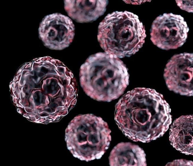
An international research team headed by Johannes Karges, PhD, of the faculty of chemistry and biochemistry at Ruhr University Bochum, Germany, has developed nanoparticles that accumulate in cancer cells and eliminate them after being photoactivated. The research team also labeled them in such a way that immune cells learn to eliminate similar cells throughout the body which could even mean undetected metastases can be treated.

The researchers presented their findings in the journal Nature Communications in an article titled, “Theranostic imaging and multimodal photodynamic therapy and immunotherapy using the mTOR signaling pathway.”

“Tumor metastases are considered the leading cause of cancer-associated deaths,” the researchers wrote. “While clinically applied drugs have demonstrated to efficiently remove the primary tumor, metastases remain poorly accessible. To overcome this limitation, herein, the development of a theranostic nanomaterial by incorporating a chromophore for imaging and a photosensitizer for treatment of metastatic tumor sites is presented. The mechanism of action reveals that the nanoparticles are able to intervene by local generation of cellular damage through photodynamic therapy as well as by systemic induction of an immune response by immunotherapy upon inhibition of the mTOR signaling pathway which is of crucial importance for tumor onset, progression, and metastatic spreading.”

A group of 12 researchers at Rice University in Houston have used 3D printing to create near-bulletproof material made out of plastic. The novel materials can withstand being shot at by bullets traveling at 5.8 kilometers per second and are highly compressible without falling apart.

Tubulanes are theoretical microscopic structures comprised of crosslinked carbon nanotubes and the researchers sought to test if they would have the same properties when scaled up enough to be 3D printed. It turns out they did.

The researchers proved this by shooting a bullet traveling at 5.8 kilometers per second through two cubes. One cube was made from a solid polymer and the other from a polymer printed with a tubulane structure.


By subscribing, you agree to our Terms of Use and Policies You may unsubscribe at any time.

Osteoarthritis (OA) is a prevalent global health concern, posing a significant and increasing public health challenge worldwide. Recently, biomaterials have emerged as a highly promising strategy for OA therapy due to their exceptional physicochemical properties and capacity to regulate pathological processes. However, there is an urgent need for a deeper understanding of the potential therapeutic applications of these biomaterials in the clinical management of diseases, particularly in the treatment of OA. In this comprehensive review, we present an extensive discussion of the current status and future prospects concerning biomaterials for OA… More.


Herein, in this review, we summarize the advanced strategies developed for enhancing OA therapy based on the biomaterials. We conducted a comprehensive literature search using relevant databases such as PubMed, Scopus, and Web of Science. The search was focused on peer-reviewed articles and research papers published within the last ten years (from 2013 to 2023). We utilized specific keywords related to biomaterials”, biomaterials” and “osteoarthritis therapy” to retrieve relevant studies. First, we provide an overview of the pathophysiology of OA and the limitations of current treatment options. Second, we explore the various types of biomaterials which have been used for OA therapy, including nanoparticles, nanofibers, and nanocomposites. Third, we highlight the advantages and challenges associated with the use of biomaterials in OA therapy, such as toxicity, biodegradation, and regulatory issues. Finally, advanced biomaterials-based OA therapies with their potential for clinical translation and emerging biomaterials directions for OA therapy are discussed.

Characteristics of Biomaterials

Nanotechnology-boosted biomaterials have attracted considerable attention in recent years as promising candidates for revolutionizing the field of therapeutics.12,13 These materials combine the unique properties of nanotechnology with the versatility and biocompatibility of biomaterials, offering numerous advantages over existing therapeutic approaches. Nanotechnology enables the precise engineering of biomaterials at the nanoscale, allowing for the encapsulation and controlled release of therapeutic agents, such as drugs and growth factors.14–17 This feature facilitates targeted and sustained drug delivery to specific sites within the body, reducing systemic side effects and enhancing treatment efficacy. In the context of OA, this targeted drug delivery can be utilized to deliver anti-inflammatory agents or disease-modifying drugs directly to affected joint tissues, promoting tissue repair and alleviating symptoms. Furthermore, biomaterials can be designed to mimic the native tissue environment, thereby enhancing their biocompatibility and reducing the risk of adverse reactions or immune responses.18 This characteristic is crucial for successful integration and long-term functionality of biomaterials in biomedical applications. Moreover, nanomaterials can facilitate tissue regeneration by stimulating cellular responses and promoting tissue growth.19 In the context of OA, biomaterials can assist in cartilage repair and regeneration, potentially slowing down disease progression and improving joint function.3 In addition, nanotechnology allows for the customization of biomaterials with a wide range of physical, chemical, and biological properties.13 This flexibility enables the development of multifunctional biomaterials that can simultaneously perform multiple tasks, such as drug delivery, imaging, and tissue regeneration. These advantages collectively contribute to their potential as innovative solutions in addressing various biomedical challenges and improving patient outcomes. In this section, we will discuss some of the key properties of biomaterials and their impact on OA treatment.

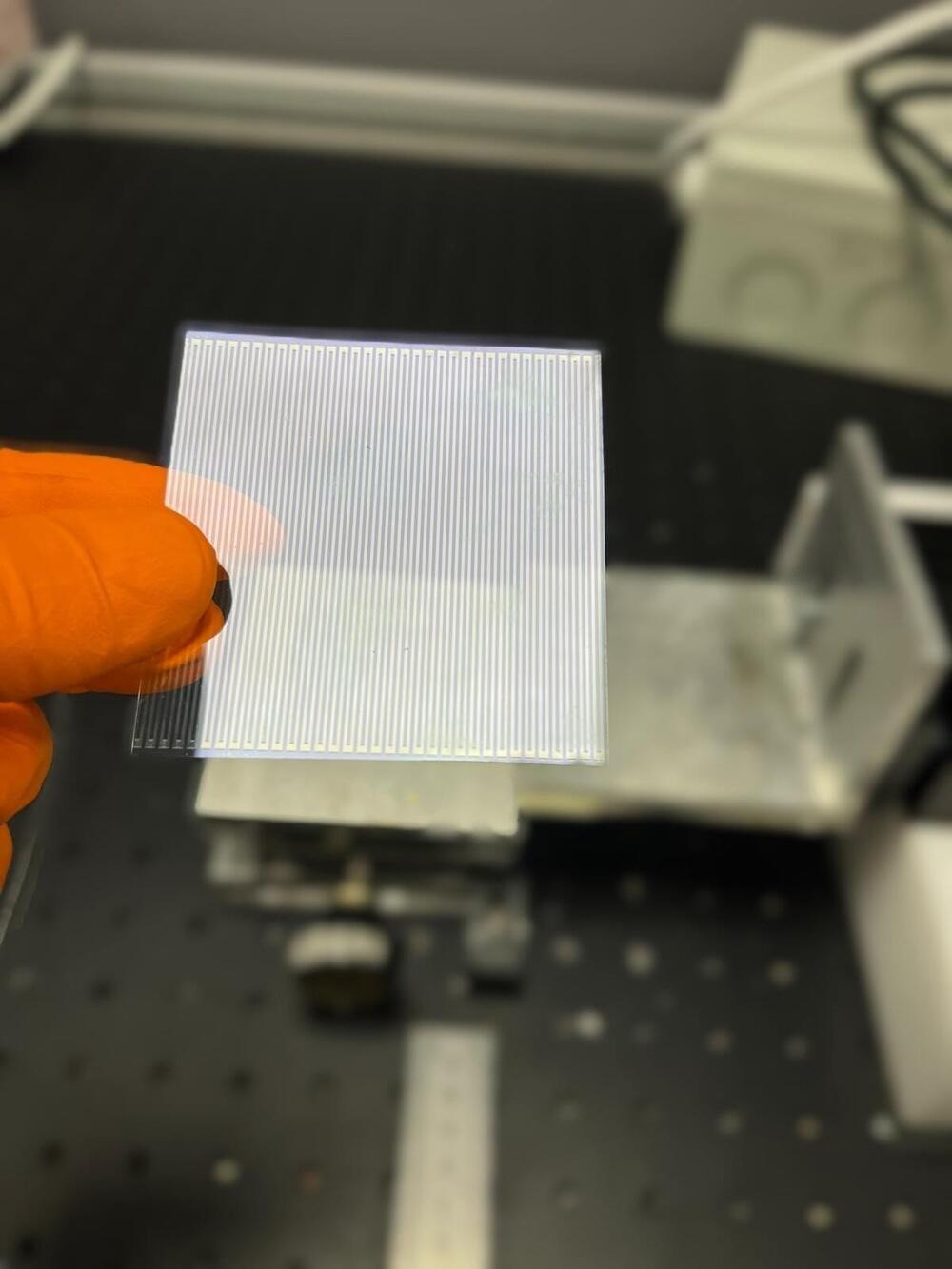
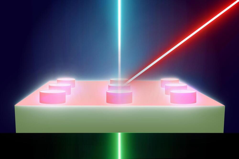
Metalens for AR and VR.


BEIJING, Sept. 8, 2023 /PRNewswire/ — WiMi Hologram Cloud Inc. (NASDAQ: WIMI) (“WiMi” or the “Company”), a leading global Hologram Augmented Reality (“AR”) Technology provider, today announced that a metasurface eyepiece for augmented reality has been developed, which is based on metasurfaces composed of artificially fabricated subwavelength structures. The metasurface eyepiece employs a special optical design and engineered anisotropic optical response to achieve an ultra-wide field of view(FOV), full-color imaging, and high-resolution near-eye display.

At the heart of the WiMi’s metalens are see-through metalens with a high numerical aperture(NA), a large area and broadband characteristics. Its anisotropic optical response allows it to perform two different optical functions simultaneously. First, it can image virtual information, acting as an imaging lens for virtual information. Second, it can transmit light, serving as a transparent glass for viewing a real-world scene. This design allows the transparent metalens to be placed directly in front of the eye without the need for additional optics, resulting in a wider FOV.

Fabrication of metalens is done using nanoimprinting technology, which is capable of fabricating large-area metalens with sub-wavelength structures. First, a mould or template with the desired structure is prepared. Then, the mould or template is contacted with a transparent substrate and the nanoscale structure is transferred by applying pressure and temperature. Through this nanoimprinting process, the subwavelength structure of the metalens is successfully replicated onto the transparent substrate, resulting in the formation of the metalens.

The ability to cure all disease slowly comes in view. ANI into Agi into ASI needs to be primary focus. Followed by genetics research, and lastly an extreme focus on Medical Nanobots. ASI will of cured most diseases by 2035–2040.


Fleets of advanced versions may one day be able to detect disease and then go about surgically treating it — without ever opening the skull.

For now, cyborgs exist only in fiction, but the concept is becoming more plausible as science progresses. And now, researchers are reporting in ACS’ Nano Letters that they have developed a proof-of-concept technique to “tattoo” living cells and tissues with flexible arrays of gold nanodots and nanowires. With further refinement, this method could eventually be used to integrate smart devices with living tissue for biomedical applications, such as bionics and biosensing.

Advances in electronics have enabled manufacturers to make integrated circuits and sensors with nanoscale resolution. More recently, laser printing and other techniques have made it possible to assemble flexible devices that can mold to curved surfaces. But these processes often use harsh chemicals, high temperatures or pressure extremes that are incompatible with living cells. Other methods are too slow or have poor spatial resolution. To avoid these drawbacks, David Gracias, Luo Gu and colleagues wanted to develop a nontoxic, high-resolution, lithographic method to attach nanomaterials to living tissue and cells.

The team used nanoimprint lithography to print a pattern of nanoscale gold lines or dots on a polymer-coated silicon wafer. The polymer was then dissolved to free the gold nanoarray so it could be transferred to a thin piece of glass. Next, the gold was functionalized with cysteamine and covered with a hydrogel layer, which, when peeled away, removed the array from the glass. The patterned side of this flexible array/hydrogel layer was coated with gelatin and attached to individual live fibroblast cells. In the final step, the hydrogel was degraded to expose the gold pattern on the surface of the cells. The researchers used similar techniques to apply gold nanoarrays to sheets of fibroblasts or to rat brains. Experiments showed that the arrays were biocompatible and could guide cell orientation and migration.

Researchers have created a new 3D-printed substance dubbed “engineered living material.”

Removing pollutants from water is a crucial and arduous process to ensure that it is free from harmful contaminants. In recent years, several approaches and technologies for water pollution remediation have been developed and employed, including filtration, nano-materials, and chemical treatment, to mention a few.

Now, researchers at the University of California, San Diego, have developed a new environmentally friendly method for removing chemical contaminants from water bodies.


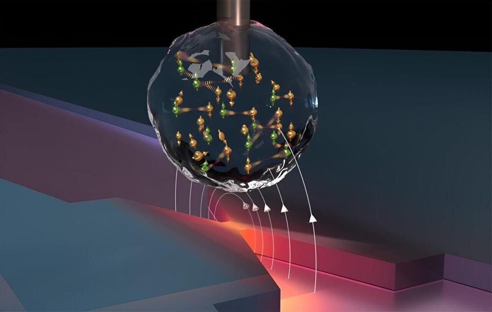
National University of Singapore (NUS) scientists demonstrated a conceptual breakthrough by fabricating atomically precise quantum antidots (QAD) using self-assembled single vacancies (SVs) in a two-dimensional (2D) transition metal dichalcogenide (TMD).

Quantum dots confine electrons on a nanoscale level. In contrast, an antidot refers to a region characterized by a potential hill that repels electrons. By strategically introducing antidot patterns (“voids”) into carefully designed antidot lattices, intriguing emerge.

These structures exhibit periodic potential modulation to change 2D electron behavior, leading to novel transport properties and unique quantum phenomena. As the trend towards miniaturized devices continue, it is important to accurately control the size and spacing of each antidot at the . This control together with resilience to environmental perturbations is crucial to address technological challenges in nanoelectronics.