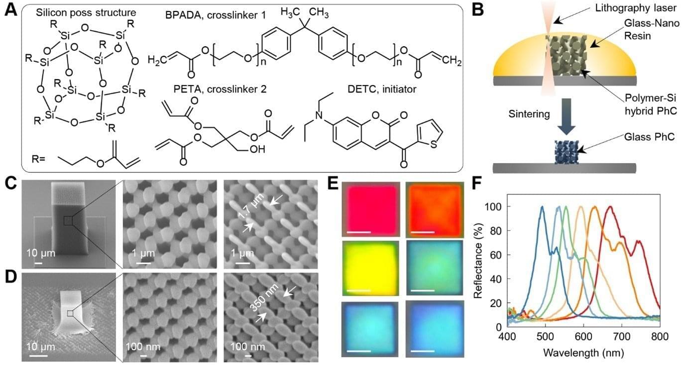
Toggle light / dark theme

Plasma-synthesized photothermal material could enable efficient solar-powered water purification

Technology for converting solar energy into thermal energy is ever evolving and has numerous applications. A breakthrough in the laboratory of Professor My Ali El Khakani at Institut national de la recherche scientifique (INRS) has made a significant contribution to the field.

Professor El Khakani specializes in plasma-laser processes for the development of nanostructured materials. He and his team at the Énergie Matériaux Télécommunications Research Center have developed a new photothermal material that converts sunlight into heat with unmatched efficiency. The results of their work were published in the journal Scientific Reports.

For several decades, stoichiometric titanium oxides have been known for their exceptional photocatalytic properties. A sub-stoichiometric form of this material, characterized by a slight deficiency in , is referred to as “Magnéli phases,” with specific compositions exhibiting distinct properties.

Scientists discover simpler is better when it comes to terahertz time-domain spectroscopy

A simple tweak to the usual setup is all that is needed to enhance a spectroscopy technique that uses waves in the terahertz region to probe samples, RIKEN physicists have discovered. The findings are published in the journal Applied Physics Letters.

Developing techniques that can obtain spectra from tiny regions extremely rapidly is the ultimate goal of a team that Norihiko Hayazawa of the RIKEN Center for Advanced Photonics belongs to.

Until recently, the scientists had been focusing on obtaining spectra from nanoscale regions on samples. But now they are concentrating on acquiring spectra very quickly—on the order of billionths of seconds (nanoseconds)—to minimize fluctuations induced by the ambient environment.

Researchers discover a new type of quantum entanglement

A study from Technion unveils a newly discovered form of quantum entanglement in the total angular momentum of photons confined in nanoscale structures. This discovery could play a key role in the future miniaturization of quantum communication and computing components.

Quantum physics sometimes leads to very unconventional predictions. This is what happened when Albert Einstein and his colleagues, Boris Podolsky and Nathan Rosen (who later founded the Faculty of Physics at Technion), found a scenario in which knowing the state of one particle immediately affects the state of the other particle, no matter how great the distance between them. Their historic 1935 paper was nicknamed EPR after its three authors (Einstein–Podolsky–Rosen).

The idea that knowing the state of one particle will affect another particle located at a huge distance from it, without physical interaction and information transfer, seemed absurd to Einstein, who called it “spooky action at a distance.”

Gold nanoclusters reveal magnetic spin’s potential role in catalytic efficiency

Recently, a team of researchers from the Hefei Institutes of Physical Science of the Chinese Academy of Sciences (CAS) consecutively removed the innermost atom and the outermost electron of a gold nanoparticle—without disturbing its overall structure. This precise manipulation allowed them to probe how the magnetic spin of the material influences its catalytic activity.

The work, led by Prof. Wu Zhikun in collaboration with Prof. Yang from the Institute of Process Engineering, CAS and Prof. Tang from Chongqing University, was published in Science Advances.

Gold nanoclusters—tiny particles composed of from a few to hundreds of —are ideal models for studying how atomic structure affects . But tuning the structure of such clusters atom by atom, especially when they’re relatively large and complex, has long been a major challenge.

Nonlinear sound-sheet microscopy

Key to this innovation in ultrasound imaging—a method called Nonlinear sound sheet microscopy —was the discovery of a sound-reflecting probe. The author said: “This probe is a nanoscale gas-filled vesicle that lights up in ultrasound images, making cells visible. These vesicles have a protein shell and we can engineer them to tune their brightness in images. We used these gas vesicles to track cancer cells.”

In addition to revealing cells, the team used ultrasound and microbubbles as probes circulating in the blood stream to detect brain capillaries. The author said: “To our knowledge, nonlinear sound sheet microscopy is the first technique capable of observing capillaries in living brains. This breakthrough has tremendous potential to diagnose small vessel diseases in patients.” Since microbubble probes are already approved for human use, this technique could be deployed in hospitals in a few years.


Ultrasound is one of the most widely used imaging techniques in medicine, but up until recently it hardly played a role in imaging the tiniest structures of our bodies such as cells. “Clinical ultrasound, like the kind used for pregnancy scans, creates real-time images of body parts”, the first author explains. “It allows diagnosis of various diseases, or to monitor a developing baby. However, what is going on at a microscopic level remains hidden.”

Now, a team of scientists managed to image specifically labelled cells in 3D with ultrasound. For the first time, they imaged living cells inside whole organs across volumes the size of a sugar cube. In comparison, current light-based microscopes often require imaging of non-living samples, the author says. “The sample or organ of interest has to be removed and processed, and you lose the ability to track activity of cells over time”

Aiming for Lighter Light Sails

Norte and his colleagues initially considered patterning the light sails with an array of identical circular holes, but such a pattern would reduce the overall effect of the powering laser. As the sail speeds up and moves away from the laser, the wavelength it preferentially reflects will shift because of the Doppler effect, and the sail will subsequently receive less of a push. What is needed instead is a pattern that can handle Doppler-shift changes while remaining highly reflective.

To find the optimal pattern, the researchers turned to a neural network, which predicted an optimal shape that is oblong rather than circular. “It looks like a potato,” says Miguel Bessa of Brown University, Rhode Island, who led the theory side of the project. Specifically, the team arranged several potato shapes in a repeating five-neighbor pattern, or pentagonal lattice. The potato-shaped arrangement allows the system to respond to a broader range of wavelengths without having to make it thicker and thus heavier.

The researchers are now working on increasing the size of their sail and looking into ways to test how well it flies. Norte notes that the light sail is just a means to accelerate the nanospacecraft, which will include a microchip, cameras, and other instruments. All those parts need to be miniaturized so that they weigh less than one gram total. “We are really trying to use nanotechnology to go faster and further than we have been able to with traditional spacecraft,” Norte says.

Combining polarized light methods reveals hidden molecular orientations with precision

Image quality often makes the difference between an amazing multimedia experience, like feeling immersed in a high-definition movie, and a visual letdown. When it comes to biomolecular imaging, the details matter even more. When scientists increase resolution in quantitative imaging, they improve accuracy and confidence in results, ultimately facilitating discoveries in studies of proteins, cells and other biomedical applications.

Scientists have long been able to look at to study their nanoscale structures and dynamics in . However, distinguishing between two closely spaced dipole emitters, which are that can emit light in specific directions and intensities, has remained a major challenge, especially when such molecules emit light at the same time and are spatially coincident, or located at nearly the same point in space.

This limitation has hindered researchers’ ability to measure the orientation and angular separation of dipoles accurately, which is vital to understanding their rotational dynamics in crowded cellular environments.

Engineers develop a way to mass manufacture nanoparticles that deliver cancer drugs directly to tumors

Polymer-coated nanoparticles loaded with therapeutic drugs show significant promise for cancer treatment, including ovarian cancer. These particles can be targeted directly to tumors, where they release their payload while avoiding many of the side effects of traditional chemotherapy.

Over the past decade, MIT Institute Professor Paula Hammond and her students have created a variety of these particles using a technique known as layer-by-layer assembly. They’ve shown that the particles can effectively combat cancer in mouse studies.

To help move these nanoparticles closer to human use, the researchers have now come up with a manufacturing technique that allows them to generate larger quantities of the particles, in a fraction of the time.

/* */