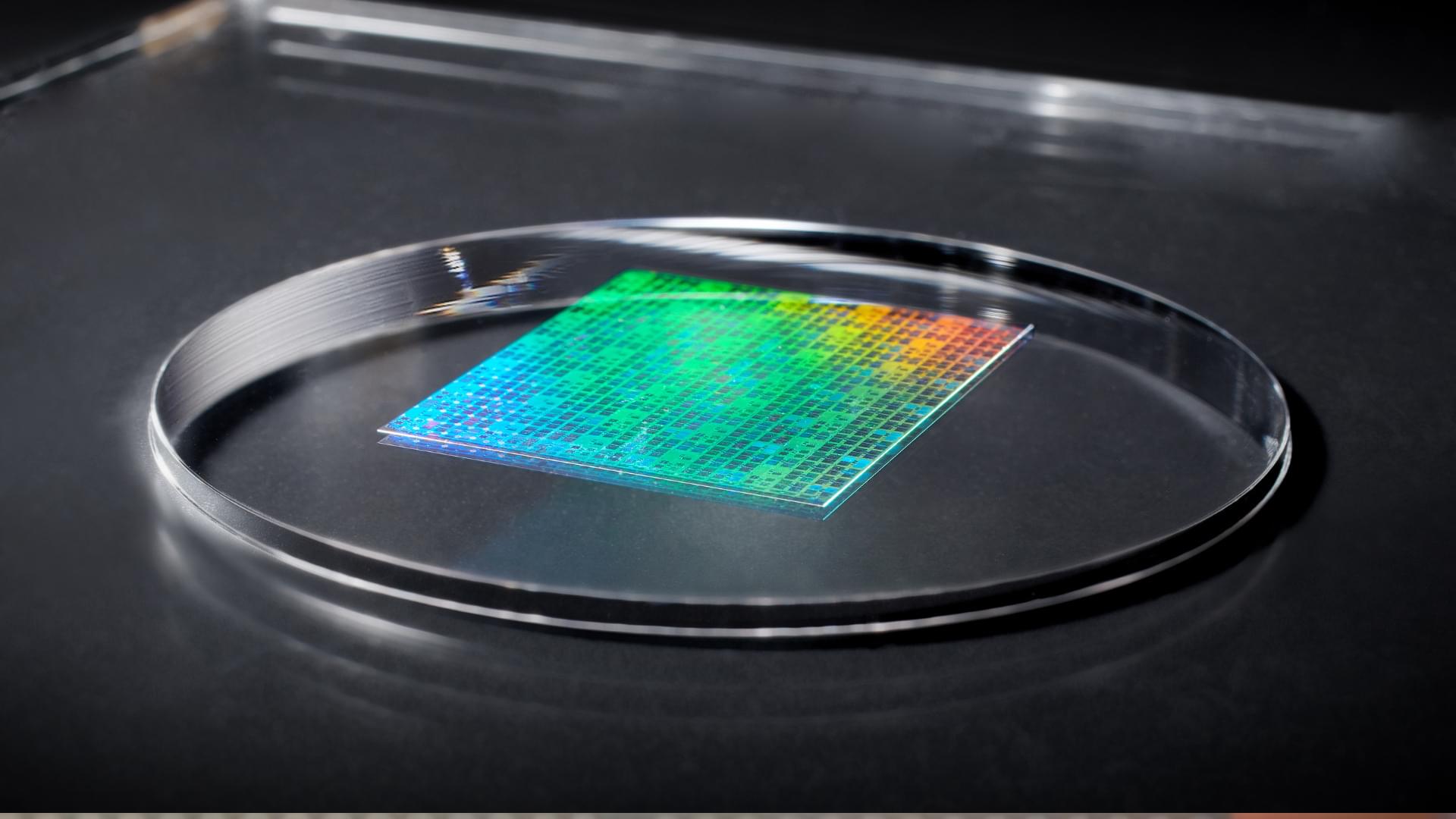
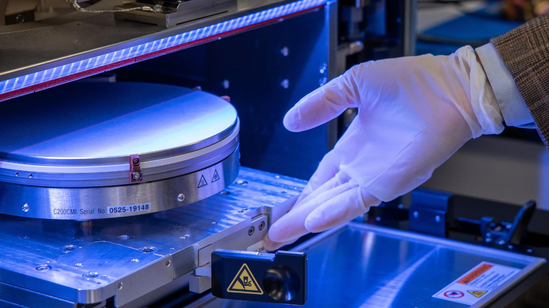
Questions remain over whether nanoimprint can shoulder even a slice of next-generation logic.
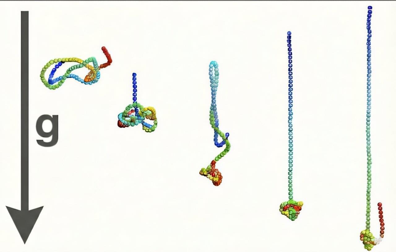
Knots are everywhere—from tangled headphones to DNA strands packed inside viruses—but how an isolated filament can knot itself without collisions or external agitation has remained a longstanding puzzle in soft-matter physics.
Now, a team of researchers at Rice University, Georgetown University and the University of Trento in Italy has uncovered a surprising physical mechanism that explains how a single filament, even one too short or too stiff to easily wrap around itself, can form a knot while sinking through a fluid under strong gravitational forces.
The discovery, published in Physical Review Letters, provides new insight into the physics of polymer dynamics, with implications ranging from understanding how DNA behaves under confinement to designing next-generation soft materials and nanostructures.
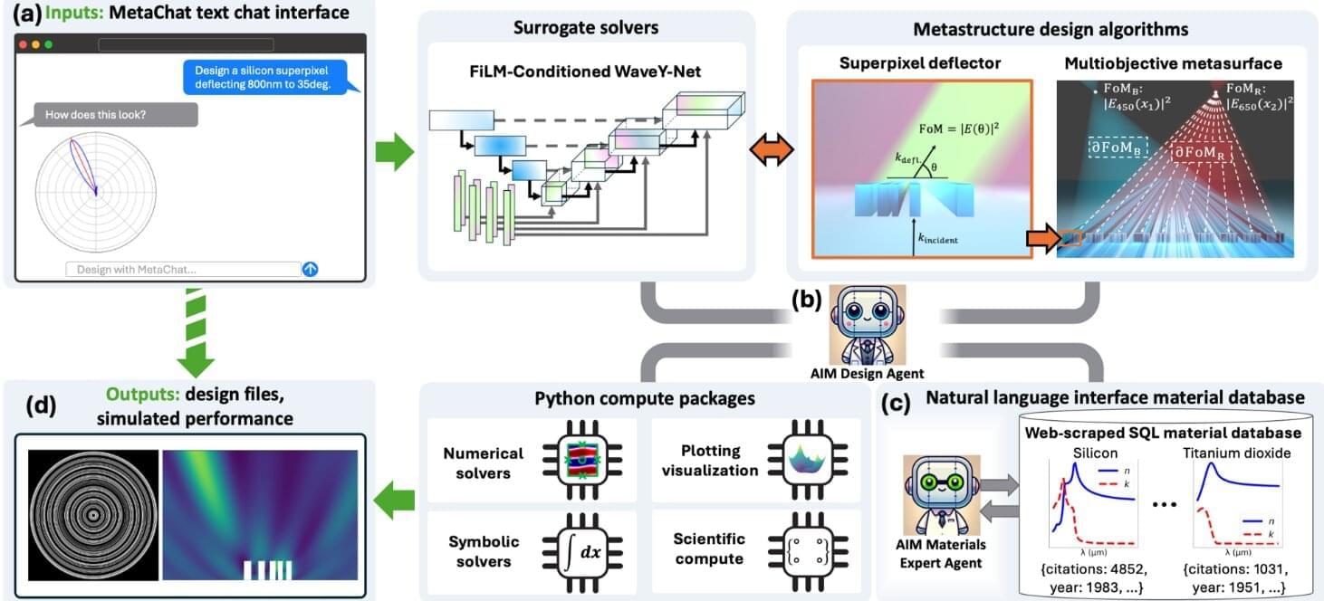
Stanford engineers debuted a new framework introducing computational tools and self-reflective AI assistants, potentially advancing fields like optical computing and astronomy.
Hyper-realistic holograms, next-generation sensors for autonomous robots, and slim augmented reality glasses are among the applications of metasurfaces, emerging photonic devices constructed from nanoscale building blocks.
Now, Stanford engineers have developed an AI framework that rapidly accelerates metasurface design, with potential widespread technological applications. The framework, called MetaChat, introduces new computational tools and self-reflective AI assistants, enabling rapid solving of optics-related problems. The findings were reported recently in the journal Science Advances.
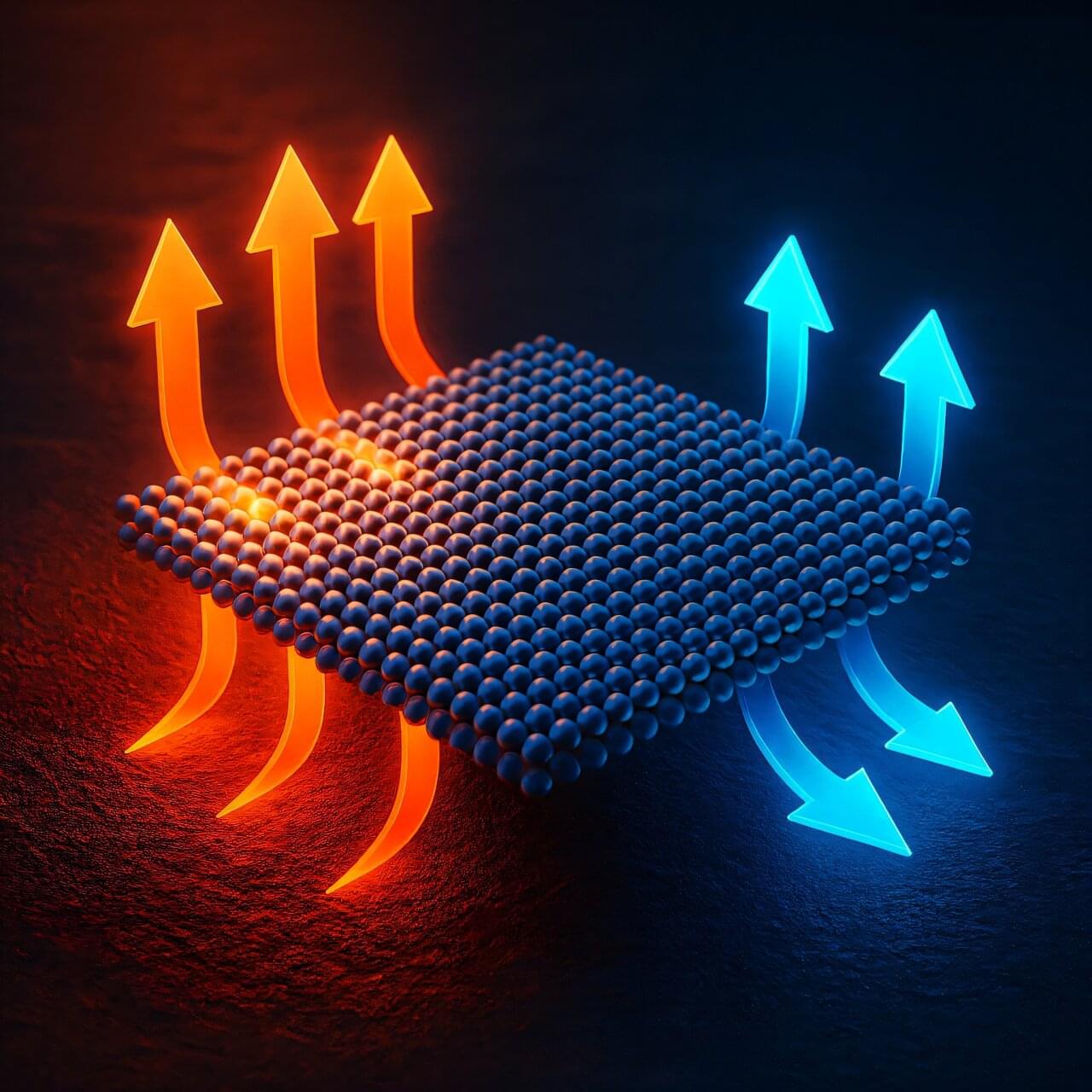
For much of my career, I have been fascinated by the ways in which materials behave when we reduce their dimensions to the nanoscale. Over and over, I’ve learned that when we shrink a material down to just a few nanometers in thickness, the familiar textbook rules of physics begin to bend, stretch, or sometimes break entirely. Heat transport is one of the areas where this becomes especially intriguing, because heat is carried by phonons—quantized vibrations of the atomic lattice—and phonons are exquisitely sensitive to spatial confinement.
A few years ago, something puzzling emerged in the literature. Molecular dynamics simulations showed that ultrathin silicon films exhibit a distinct minimum in their thermal conductivity at around one to two nanometers thickness, which corresponds to just a few atomic layers. Even more surprisingly, the thermal conductivity starts to increase again if the material is made even thinner, approaching extreme confinement and the 2D limit.
This runs counter to what every traditional model would predict. According to classical theories such as the Boltzmann transport equation or the Fuchs–Sondheimer boundary-scattering framework, reducing thickness should monotonically suppress thermal conductivity because there is simply less room for phonons to travel freely and carry heat around. Yet the simulations done by the team of Alan McGaughey at Carnegie Mellon University in Pittsburgh insisted otherwise, and no established theory could explain why.
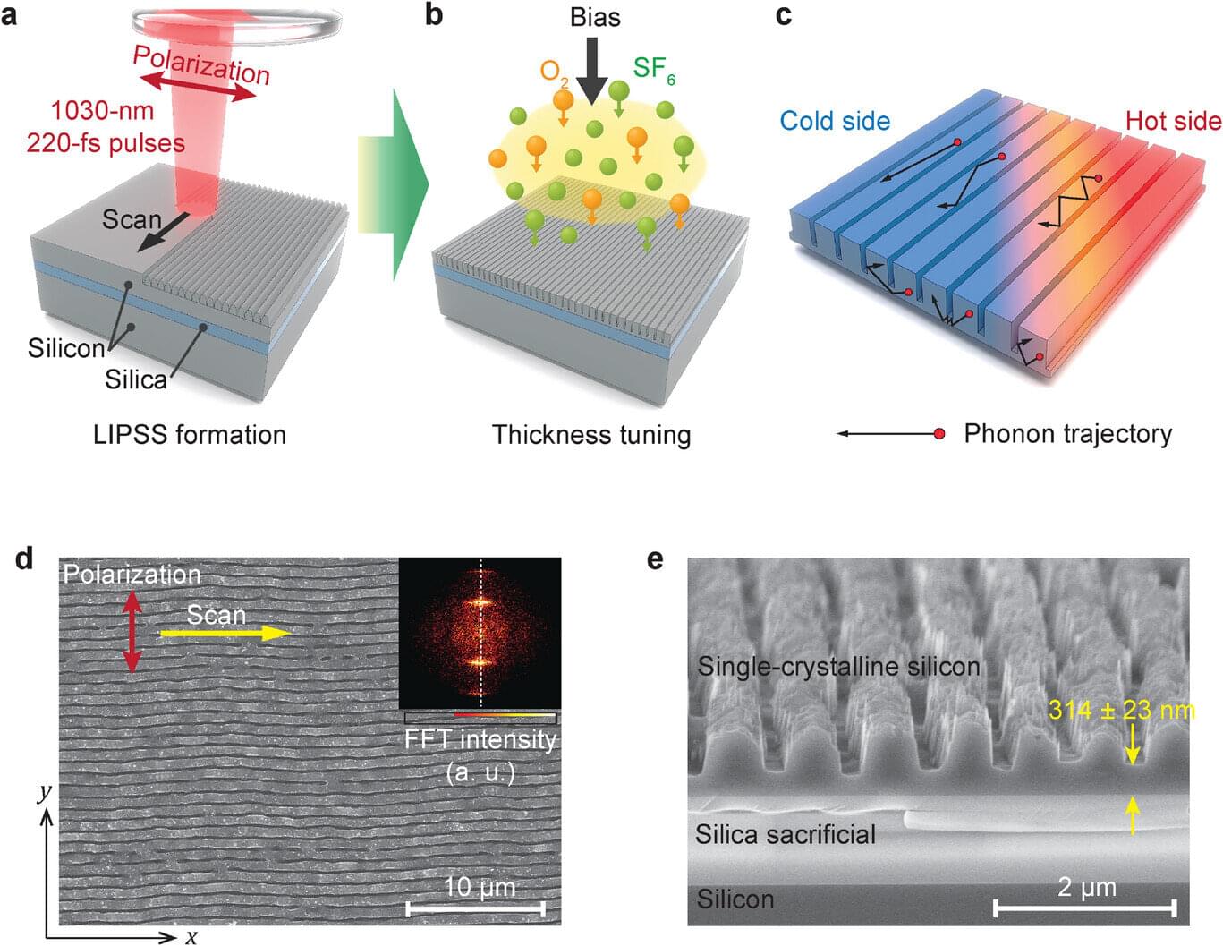
Femtosecond laser-induced periodic surface structures can be used to control thermal conductivity in thin film solids, report researchers from Japan. Their innovative method, which leverages high-speed laser ablation, produces parallel nanoscale grooves with unprecedented throughput that is 1,000 times stronger than conventional approaches, strategically altering phonon scattering in the material.
This scalable and semiconductor-ready approach could make it possible to mass-produce thermal engineering structures while maintaining laboratory-level precision.
This case-control study found that adults with schizophrenia had significantly greater frontal cortex serotonin release than healthy controls, and greater release correlated with more severe negative symptoms.
Question Is serotonin release altered in vivo in schizophrenia, and is it associated with negative symptom severity?
Findings In this case-control neuroimaging study that included 54 adults, frontal cortex serotonin release was significantly greater in the 26 people with DSM-5 schizophrenia compared with 28 matched healthy controls. In schizophrenia, greater frontal cortex serotonin release was associated with more severe baseline negative symptoms.
Meaning Findings suggest that serotonergic dysfunction in the pathophysiology of schizophrenia was associated with negative symptoms, identifying the regulation of serotonergic neurotransmission as a potential target to treat negative symptoms.
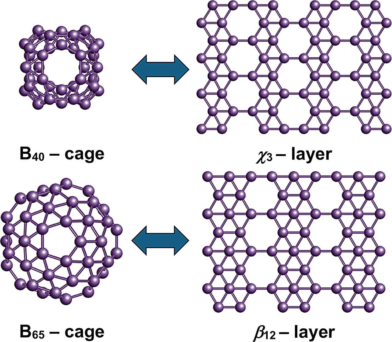
Boron, a chemical element next to carbon in the periodic table, is known for its unique ability to form complex bond networks. Unlike carbon, which typically bonds with two or three neighboring atoms, boron can share electrons among several atoms. This leads to a wide variety of nanostructures. These include boron fullerenes, which are hollow, cage-like molecules, and borophenes, ultra-thin metallic sheets of boron atoms arranged in triangular and hexagonal patterns.
Dr. Nevill Gonzalez Szwacki has developed a model explaining the variety of boron nanostructures. The analysis, published in the journal 2D Materials, combines more than a dozen known boron nanostructures, including the experimentally observed B₄₀ and B₈₀ fullerenes.
Using first-principles quantum-mechanical calculations, the study shows that the structural, energetic, and electronic properties of these systems can be predicted by looking at the proportions of atoms with four, five, or six bonds. The results reveal clear links between finite and extended boron structures. The B₄₀ cage corresponds to the χ₃ borophene layer, while B₆₅, B₈₀, and B₉₂ connect with the β₁₂, α, and bt borophene sheets, respectively. These structural links suggest that new boron cages could be created by using known two-dimensional boron templates.
Molybdenum disulfide MoS2 is a groundbreaking material for electronics applications. As a two-dimensional layer similar to graphene, it is an excellent semiconductor, and can even become intrinsically superconducting under the right conditions. It’s not particularly surprising that science fiction authors have already been speculating about molycircs, fictional computer circuits built from MoS2, for years—and that physicists and engineers are directing huge research efforts at this material.
Researchers at the University of Regensburg, have many years of expertise with diverse quantum materials—in particular also with carbon nanotubes, tube-like macromolecules made from carbon atoms alone.
“It was an obvious next step to now focus on MoS2 and its fascinating properties,” said Dr. Andreas K. Hüttel, head of the research group Nanotube Electronics and Nanomechanics in Regensburg. In cooperation with Prof. Dr. Maja Remškar, Jožef Stefan Institut Ljubljana, a specialist in the crystalline growth of molybdenum disulfide nanomaterials, his research group started working on quantum devices based on MoS2 nanotubes.