Toggle light / dark theme

Viral Appropriation of Specificity Protein 1 (Sp1): The Role of Sp1 in Human Retro- and DNA Viruses in Promoter Activation and Beyond

Specificity protein 1 (Sp1) is a highly ubiquitous transcription factor and one employed by numerous viruses to complete their life cycles. In this review, we start by summarizing the relationships between Sp1 function, DNA binding, and structural motifs. We then describe the role Sp1 plays in transcriptional activation of seven viral families, composed of human retro- and DNA viruses, with a focus on key promoter regions. Additionally, we discuss pathways in common across multiple viruses, highlighting the importance of the cell regulatory role of Sp1. We also describe Sp1-related epigenetic and protein post-translational modifications during viral infection and how they relate to Sp1 binding. Finally, with these insights in mind, we comment on the potential for Sp1-targeting therapies, such as repurposing drugs currently in use in the anti-cancer realm, and what limitations such agents would have as antivirals.

Automated chloroplast screening platform speeds up crop trait development

Chloroplasts—the “light power plants” of plant cells—are increasingly the focus of synthetic biology. These organelles house the photosynthetic apparatus and host several metabolic pathways that are of great interest for engineering new traits. Gene insertion into chloroplasts is precise and carries a lower risk of transgene escape.

Despite this potential, chloroplast biotechnology remains in its infancy because standardized, scalable methods for rapid testing of diverse genetic parts have been missing. A research team from the Max Planck Institute for Terrestrial Microbiology in Marburg has now presented a micro‑algal platform that allows automated, fast, and large‑scale testing of chloroplast genetic modifications.

The study is published in the journal Nature Plants.

Small brain region linked to schizophrenia risk through unique gene changes

New research published in the American Journal of Psychiatry provides new molecular insights into the role of the habenula, a pea-sized brain region that helps regulate motivation and mood, in contributing to the risk of schizophrenia. A team of researchers at Lieber Institute for Brain Development and Johns Hopkins found that many schizophrenia-related molecular changes appear to be specific to this region, suggesting the habenula could be a potential target for future treatments.

Schizophrenia is a heritable disorder, and a combination of multiple genetic variants contributes to it. This study sought to understand how molecular changes in the habenula region of the brain contribute to the development of . The authors note that they focused on the habenula because of its “emerging role in and functional influence on neurotransmitter systems impacted in schizophrenia.”

The study team, led by Ege A. Yalcinbas, Ph.D., used cutting-edge molecular techniques to analyze postmortem human brains, resulting in the creation of the first cell-by-cell and within-cell gene expression map of the human habenula (Hb). They then compared from 35 individuals with schizophrenia and 33 nonpsychiatric donors.

Functionally dominant hotspot mutations of mitochondrial ribosomal RNA genes in cancer

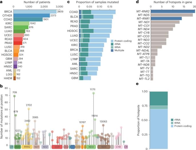
To study selection for somatic single nucleotide variants (SNVs) in tumor mtDNA, we identified somatic mtDNA variants across primary tumors from the GEL cohort (n = 14,106). The sheer magnitude of the sample size in this dataset, in conjunction with the high coverage depth of mtDNA reads (mean = 15,919×), enabled high-confidence identification of mtDNA variants to tumor heteroplasmies of 5%. In total, we identified 18,104 SNVs and 2,222 indels (Supplementary Table 1), consistent with previously reported estimates of approximately one somatic mutation in every two tumors1,2,3. The identified mutations exhibited a strand-specific mutation signature, with a predominant occurrence of CT mutations on the heavy strand and TC on the light strand in the non-control region that was reversed in the control region2 (Extended Data Fig. 1a, b). These mutations occur largely independently of known nuclear driver mutations, with the exception of a co-occurrence of TP53 mutation and mtDNA mutations in breast cancer (Q = 0.031, odds ratio (OR) = 1.43, chi-squared test) (Extended Data Fig. 2a and Supplementary Table 4).

Although the landscape of hotspot mutations in nuclear-DNA-encoded genes is relatively well described, a lack of statistical power has impeded an analogous, comprehensive analysis in mtDNA16,17. To do so, we applied a hotspot detection algorithm that identified mtDNA loci demonstrating a mutation burden in excess of the expected background mutational processes in mtDNA (Methods). In total, we recovered 138 unique statistically significant SNV hotspots (Q 0.05) across 21 tumor lineages (Fig. 1a, b and Supplementary Table 2) and seven indel hotspots occurring at homopolymeric sites in complex I genes, as previously described by our group (Extended Data Fig. 2b and Supplementary Table 3). SNV hotspots affected diverse genetic elements, including protein-coding genes (n = 96 hotspots, 12 of 13 distinct genes), tRNA genes (n = 8 hotspots, 6 of 22 distinct genes) and rRNA genes (n = 34 hotspots, 2 of 2 genes) (Fig. 1b, c, e).

Holographic optogenetics could enable faster brain mapping for new discoveries

Recent technological advances have opened new possibilities for neuroscience research, allowing researchers to map the brain’s structure and synaptic connectivity (i.e., the junctions via which neurons communicate with each other) with increasing precision.

Despite these developments, most widely employed methods to image synaptic connectivity are slow and fail to precisely record changes in the connections between in vivo, or in other words, while animals are awake and engaging in specific activities.

Two different research groups, one based at Columbia University and UC Berkeley, and the other at the Vision Institute of Sorbonne University in Paris, introduced a promising approach to study synapses in vivo. Their proposed mapping strategies, outlined in two Nature Neuroscience papers, combine holographic optogenetics, a method to selectively and precisely stimulate or silence specific neuron populations, with .

A gene from 100-year-olds could help kids who age too fast

Scientists have uncovered a breakthrough in the fight against a rare genetic condition that causes children to age much faster than normal. The discovery involves “longevity genes” found in people who live exceptionally long lives, often beyond 100 years. Researchers from the University of Bristol and IRCCS MultiMedica found that these genes, which help maintain the health of the heart and blood vessels during aging, could reverse some of the damage caused by this devastating disease.

The study, published in Signal Transduction and Targeted Therapy, is the first to show that a gene from long-lived individuals can slow down heart aging in a model of Progeria. Known scientifically as Hutchinson-Gilford Progeria Syndrome (HGPS), this rare and fatal disorder causes children to exhibit signs of “accelerated aging.”

Progeria stems from a mutation in the LMNA gene, which leads to the creation of a harmful protein called progerin. This protein disrupts normal cell function, particularly in the heart and blood vessels. Most affected children die in their teenage years from heart complications, though some, like Sammy Basso — the oldest known person with Progeria — live longer. Sammy passed away on October 24, 2024, at the age of 28.

HUMAN Stem Cells Have Reversed Age In Monkey’s with ZERO Side Effects

Age Reversal in Primates has been achieved. We have it now.

Anti-aging gene therapy, stem cell rejuvenation, and FOXO3 longevity research take center stage in this episode of Longevity Science News with Emmett Short. This groundbreaking study out of Beijing shows that gene-edited human stem cells—specifically FOXO3-enhanced senescence-resistant mesenchymal progenitor cells (SRCs)—can reverse biological aging in elderly monkeys, restoring youthful brain structure, bone density, immune strength, and even ovarian function. By upgrading the FOXO3 longevity gene, scientists created stem cells that resist cellular senescence, DNA damage, and oxidative stress, effectively making the monkeys younger from the inside out. MRI scans revealed increased cortical thickness and improved memory-related connectivity, while biological age clocks showed a 3–5 year reversal across 54% of tissues—equivalent to 9–15 years of human rejuvenation. Emmett explains how these anti-aging stem cells, epigenetic resets, and exosome-based rejuvenation pathways could revolutionize regenerative medicine, longevity biotech, and future human trials. He also explores the costs, ethics, and long-term implications of turning back time at the cellular level. If you’re passionate about biohacking, gene editing, lifespan extension, or the future of anti-aging science, this is the video for you.

HUME BODY ANALYZER:
Use Code: LONGEVITY for up to 50% OFF
https://humehealth.com//discount/LONGEVITY?redirect=/pages/h…=LONGEVITY

Watch next: Artificial Blood: The SciFi Anti-Aging Tech That’s Now in Human Trials.
https://www.youtube.com/watch?v=3xz1lcGdQPc.

If you’re into the cutting edge of anti-aging science, subscribe and share with someone who wants to live longer, healthier, and stronger.

⏱️ Chapters:

Merge Labs, Sam Altman, genetic targeting, ultrasound systems, Open AI, mechanosensitive channels

In recent years, neuroengineers have devised a number of new modalities for interfacing with the nervous system. Among these are optical stimulation, vibrational stimulation, and optogenetics. A newer and perhaps more promising technology is sonogenetics.

Sonogenetics, the use of focused ultrasound to control cells that have been made ultrasound-responsive via gene delivery, is moving from compelling papers to a potential platform strategy. From a neurotech commercialization standpoint, the significance of sonogenetics is less about a single lab trick and more about the emerging convergence of three capabilities: precise genetic targeting, durable and safe delivery, and field-robust ultrasound systems that work the first time outside the origin lab.

One commercial firm that may be exploiting this technology is Merge Labs. The startup recently made a big splash with a $250 million investment from Open AI and Sam Altman. While the company has not yet released its website and the technical personnel behind the company have not been identified, it is rumored to be working with focused ultrasound implants and sonogenetics as gene therapy. If Merge and its peers can validate durable expression, predictable dose–response, and reliable outside-the-lab bring-up, a first wave of indications will likely sit at the intersection of neurology, psychiatry, and rehabilitation, with longer-term spillover into human-machine interaction.

Imaging study shows how brains go off-track in rare childhood disorder

Researchers at the VIB-UAntwerp Center for Molecular Neurology have visualized how brain network development is altered in a model of KCNQ2-related developmental and epileptic encephalopathy, a rare childhood brain disorder. Using longitudinal imaging techniques, the team observed differences in how brain regions communicate and connect, long before behavioral symptoms appear.

KCNQ2-related developmental and epileptic encephalopathy (KCNQ2-DEE) is a rare but severe neurological disorder that affects newborns. Children with this condition typically develop seizures within days after birth and continue to face learning and movement difficulties. The disorder is caused by mutations in a potassium-channel gene that disrupts normal brain activity.

To investigate how this disorder affects , the team of Professor Sarah Weckhuysen visualized and structure throughout early growth in mice carrying the same genetic defect. The study is published in the journal eBioMedicine.

Another protease, pepsin, cuts in the same general region of the antibody molecule as papain but on the carboxy-terminal side of the disulfide bonds (see Fig

3.3). This produces a fragment, the F(ab′)2 fragment, in which the two -binding arms of the antibody molecule remain linked. In this case the remaining part of the is cut into several small fragments. The F(ab′)2 fragment has exactly the same antigen-binding characteristics as the original antibody but is unable to interact with any effector molecule. It is thus of potential value in therapeutic applications of antibodies as well as in research into the functional role of the Fc portion.

Genetic engineering techniques also now permit the construction of many different -related molecules. One important type is a truncated Fab comprising only the of a linked by a stretch of synthetic peptide to a V domain of a . This is called , named from Fragment v ariable. Fv molecules may become valuable therapeutic agents because of their small size, which allows them to penetrate tissues readily. They can be coupled to protein toxins to yield immunotoxins with potential application, for example, in tumor therapy in the case of a Fv specific for a tumor (see Chapter 14).

/* */