The development of biomaterials for artificial organs and tissues is an active area of research due to increases in accidental injuries and chronic diseases, along with the entry into a super-aged society. 3D bioprinting technology, which uses cells and biomaterials to create three-dimensional artificial tissue structures, has recently gained popularity. However, commonly used hydrogel-based bioinks can cause cytotoxicity due to the chemical crosslinking agent and ultraviolet light that connect the molecular structure of photocuring 3D-printed bioink.
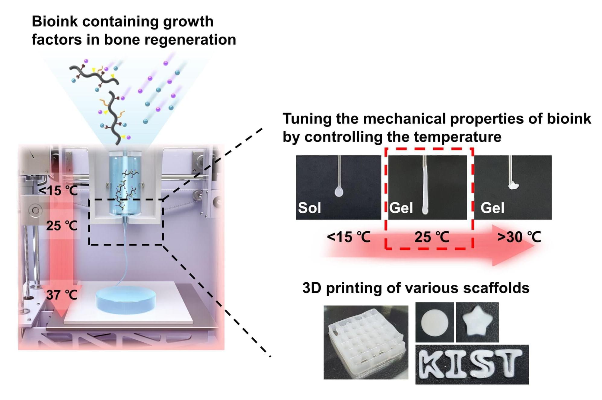
Dr. Song Soo-chang’s research team at the Center for Biomaterials, Korea Institute of Science and Technology (KIST), revealed the first development of poly(organophosphazene) hydrogel-based temperature-sensitive bioink that stably maintained its physical structure by temperature control only without photocuring, induced tissue regeneration, and then biodegraded in the body after a certain period of time.
Current hydrogel-based bioinks must go through a photocuring process to enhance the mechanical properties of the 3D scaffold after printing, with a high risk of adverse effects in the human body. In addition, there has been a possibility of side effects when transplanting externally cultured cells within bioink to increase the tissue regeneration effect.