Toggle light / dark theme

Circa 2020 Immortality of the heart and heart regeneration.


Cardiovascular diseases represent the major cause of morbidity and mortality worldwide. Multiple studies have been conducted so far in order to develop treatments able to prevent the progression of these pathologies. Despite progress made in the last decade, current therapies are still hampered by poor translation into actual clinical applications. The major drawback of such strategies is represented by the limited regenerative capacity of the cardiac tissue. Indeed, after an ischaemic insult, the formation of fibrotic scar takes place, interfering with mechanical and electrical functions of the heart. Hence, the ability of the heart to recover after ischaemic injury depends on several molecular and cellular pathways, and the imbalance between them results into adverse remodeling, culminating in heart failure. In this complex scenario, a new chapter of regenerative medicine has been opened over the past 20 years with the discovery of induced pluripotent stem cells (iPSCs). These cells share the same characteristic of embryonic stem cells (ESCs), but are generated from patient-specific somatic cells, overcoming the ethical limitations related to ESC use and providing an autologous source of human cells. Similarly to ESCs, iPSCs are able to efficiently differentiate into cardiomyocytes (CMs), and thus hold a real regenerative potential for future clinical applications. However, cell-based therapies are subjected to poor grafting and may cause adverse effects in the failing heart. Thus, over the last years, bioengineering technologies focused their attention on the improvement of both survival and functionality of iPSC-derived CMs. The combination of these two fields of study has burst the development of cell-based three-dimensional (3D) structures and organoids which mimic, more realistically, the in vivo cell behavior. Toward the same path, the possibility to directly induce conversion of fibroblasts into CMs has recently emerged as a promising area for in situ cardiac regeneration. In this review we provide an up-to-date overview of the latest advancements in the application of pluripotent stem cells and tissue-engineering for therapeutically relevant cardiac regenerative approaches, aiming to highlight outcomes, limitations and future perspectives for their clinical translation.

Cardiovascular diseases represent the major cause of morbidity and mortality worldwide, accounting for 31% of all deaths (Organization WH 2016). Myocardial infarction (MI) is associated with necrosis of the cardiac tissue due to the occlusion of the coronary arteries, a condition that irrevocably diminishes oxygen and nutrient delivery to the heart (Thygesen et al., 2007). While effective therapies, including surgical approaches, are currently used to treat numerous cardiac disorders, such as valvular or artery diseases, available therapeutic treatments for the damaged myocardium are still very limited and poorly effective. Furthermore, after an ischaemic insult, the formation of fibrotic scar takes place, interfering with mechanical and electrical functions of the cardiac tissue (Talman and Ruskoaho, 2016).

A few years ago, Jürgen Knoblich and his team at the Institute of Molecular Biotechnology of the Austrian Academy of Sciences (IMBA) have pioneered brain organoid technology. They developed a method for cultivating three-dimensional brain-like structures, so called cerebral organoids, in a dish. This discovery has tremendous potential as it could revolutionize drug discovery and disease research. Their lab grown organ-models mimic early human brain development in a surprisingly precise way, allowing for targeted analysis of human neuropsychiatric disorders, that are otherwise not possible. Using this cutting-edge methodology, research teams around the world have already revealed new secrets of human brain formation and its defects that can lead to microcephaly, epilepsy or autism.

In a new study published in Nature Biotechnology, scientists from Cambridge and Vienna present a new method that combines the organoid method with bioengineering. The researchers use special polymer fibers made of a material called PLGA) to generate a floating scaffold that was then covered with human cells. By using this ground-breaking combination of engineering and stem cell culture, the scientists are able to form more elongated organoids that more closely resemble the shape of an actual human embryo. By doing so, the organoids become more consistent and reproducible.

“This study is one of the first attempts to combine organoids with bioengineering. Our new method takes advantage of and combines the unique strengths of each approach, namely the intrinsic self-organization of organoids and the reproducibility afforded by bioengineering. We make use of small microfilaments to guide the shape of the organoids without driving tissue identity, ” explains Madeline Lancaster, group leader at MRC Laboratory of Molecular Biology in Cambridge and first author of the paper.

Materials scientists and bioengineers at the intersection of regenerative medicine and bioinspired materials seek to develop shape-programmable artificial muscles with self-sensing capabilities for applications in medicine. In a new report now published in Science Advances, Haoran Liu and a team of researchers in systems and communications engineering at the Frontier Institute of Science and Technology, Jiaotong University, China, were inspired by the coupled behavior of muscles, bones, and nerve systems of mammals and other living organisms to create a multifunctional artificial muscle in the lab. The construct contained polydopamine-coated liquid crystal elastomer (LCE) and low-melting point alloys (LMPA) in a concentric tube or rod. While the team adopted the outer liquid crystal-elastomer to mimic reversible contraction and recovery, they implemented the inner low-melting point alloy for deformation locking and to detect resistance mechanics, much like bone and nerve functions, respectively. The artificial muscle demonstrated a range of performances, including regulated bending and deformation to support heavy objects, and is a direct and effective approach to the design of biomimetic soft devices.

Soft robotics inspired by the skeleton–muscle–nerve system

Scientists aim to implement biocompatibility between soft robotic elements and human beings for assisted movement and high load-bearing capacity; however, such efforts are challenging. Most traditional robots are still in use in industrial, agricultural and aerospace settings for high-precision sensor-based, load-bearing applications. Several functional soft robots contrastingly depend on materials to improve the security of human-machine interactions. Soft robots are therefore complementary to hard robots and have tremendous potential for applications. Biomimetic constructs have also provided alternative inspiration to emulate the skeleton-muscle-nerve system to facilitate agile movement and quick reaction or thinking, with a unique body shape to fit tasks and perform diverse physiological functions. In this work, Liu et al were inspired by the fascinating idea of biomimicry to develop multifunctional artificial muscles for smart applications.

As they grow, solid tumors surround themselves with a thick, hard-to-penetrate wall of molecular defenses. Getting drugs past that barricade is notoriously difficult. Now, scientists at UT Southwestern have developed nanoparticles that can break down the physical barriers around tumors to reach cancer cells. Once inside, the nanoparticles release their payload: a gene editing system that alters DNA inside the tumor, blocking its growth and activating the immune system.

The new , described in Nature Nanotechnology, effectively stopped the growth and spread of ovarian and liver tumors in mice. The system offers a new path forward for the use of the gene editing tool known as CRISPR-Cas9 in , said study leader Daniel Siegwart, Ph.D., Associate Professor of Biochemistry at UT Southwestern.

“Although CRISPR offers a new approach for treating , the technology has been severely hindered by the low efficiency of delivering payloads into tumors,” said Dr. Siegwart, a member of the Harold C. Simmons Comprehensive Cancer Center.

Superhumans are coming! Various technological advances in the field of medicine through AI and CRISPR are going to radically alter our understanding of what it means to be human. AI and Crispr technology have been making revolutionary changes to the field of medicine. Artificial intelligence is being applied in identification of harmful genes and treatment of disease.

Multiple new gene editing technologies in addition to artificial intelligence will cause major changes in healthcare.
The gene-editing tool CRISPR, short for clustered regularly interspaced short palindromic repeats, could help us to reprogram life. It gives scientists more power and precision than they have ever had to alter human DNA.

Could it be conceivable that at one point in the future we could increase the average IQ of the population?
CRISPR technology in the future could enable efficient and affordable treatment of many diseases. It will be also probably used to enhance our intelligence.

CRISPR was originally designed as a technique for editing the DNA of living organisms, but its uses and capabilities have expanded substantially. It is now used widely in biomedical research, including research into human genetics. One of the main issues concerning technologies such as CRISPR-Cas9 is accuracy and safety, as mistakes can have significant and dangerous consequences when it comes to an individual’s genome.

David Sinclair explains the mind boggling breakthroughs of medicine in recent times.

The possibility of altering the sequences of genes and correcting the mistakes that were previously irreversible opens many new possibilities for research. Nevertheless, for now, genetic editing technology is still incapable of making the hundreds of changes presumably necessary to make a superintelligent human. But the corresponding ethical issues deserve serious attention before these capabilities become a reality.

Artificial Intelligence is touching almost every aspect of our lives. It’s reasonable to expect AI influence will only increase in the future. One of many fields heavily influenced by AI is the military. Particularly in the development of Supersoldiers. The notion of super-soldiers enhanced with biotechnology and cybernetics was once only possible in the realm of science fiction. But it may not be too long before these concepts become a reality.

A new worldwide arms race is pitting countries against each other to be the first to successfully create real genetically modified super soldiers by using tools such as CRISPR. Understandably many of these human enhancement technologies raise health and safety questions and it is more likely these enhancements will first gain traction in countries that do not place as much weight on ethical concerns.

According to US Intelligence, China has conducted “human testing” on members of the People’s Liberation Army in hope of developing soldiers with “biologically enhanced capabilities.

This has made the U.S. military’s top intelligence agencies increasingly worried but the Pentagon has significantly invested in its own research in AI and in the extension of the human senses beyond their current physical limitations, to provide soldiers with superhuman abilities.

The basics of brain-machine interfaces with AI are being developed for the military, and if the results are as successful as scientists hope they will be, soldiers could one day be enhanced with cybernetics, effectively becoming trans-human soldiers.

The US Military is also examining newly scientific tools, like genetic engineering, brain chemistry, and shrinking robotics, for even more dramatic enhancements. But most of this advanced technology remains classified.

A long-running human trial has shown that CRISPR gene editing could prove to be a highly effective way of treating serious conditions.

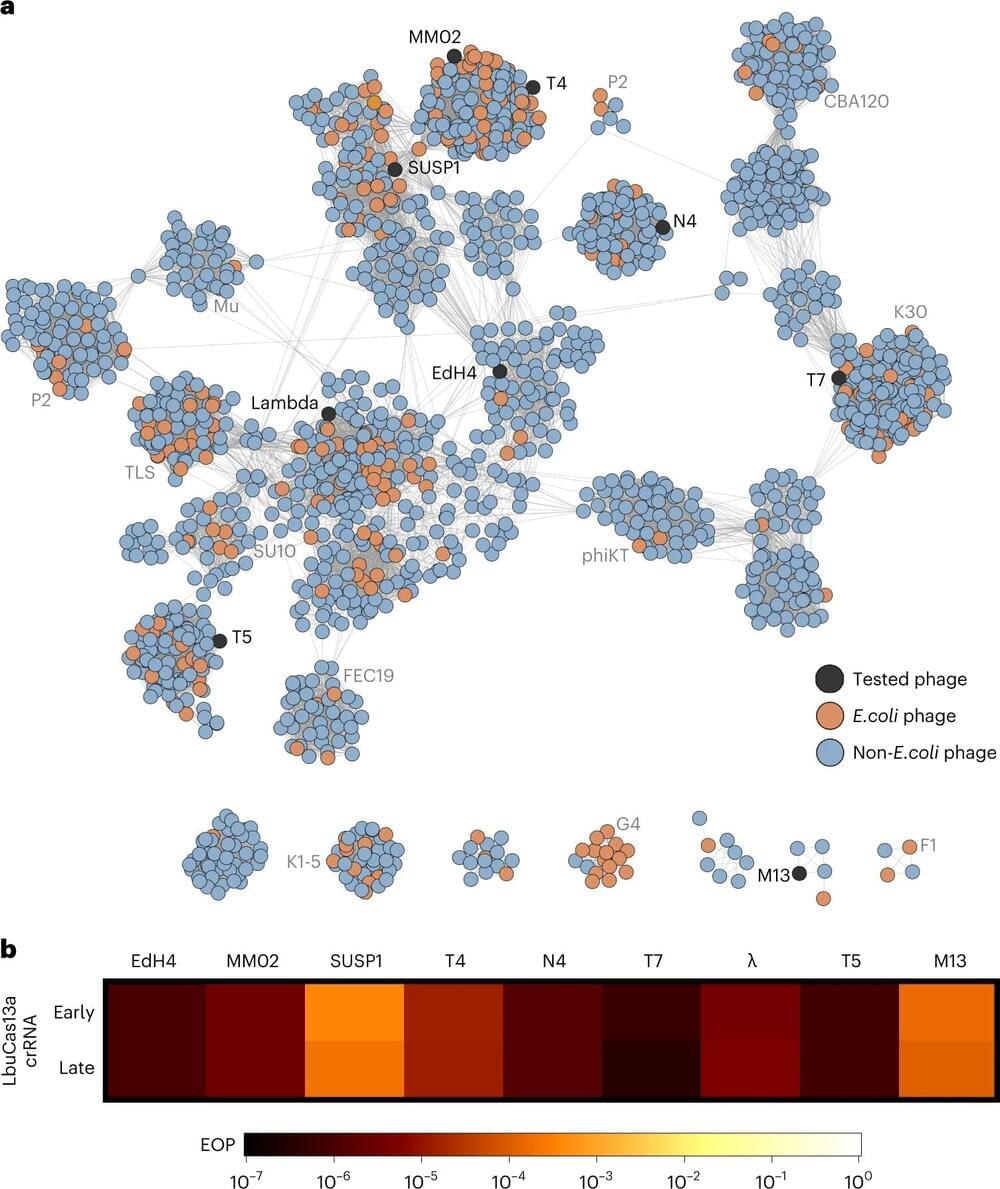
The trial, which was kicked off in 2019 by an international team of scientists, found that a new gene-editing therapy called exagamglogene autotemcel, or ex-cel for short, was able to essentially “cure” patients with transfusion-dependent beta thalassemia (TDT) or severe sickle cell disease (SCD), two blood disorders that are conventionally treated using blood transfusions.

It’s a promising new use of the technology. Around 100,000 Americans are affected by TDT, while SCD affects an estimated 300 to 3,000. And in a broader sense, the results suggest that tinkering with genetic code could come to be a practical, widespread new area of medicine.

Tel Aviv University researchers have published a new study in Nature outlining how a type of white blood cell can be engineered to secrete anti-human immunodeficiency virus (HIV) antibodies. Based on the results of this study, the team are hopeful that they will be able to produce a one-time medication for acquired immune deficiency syndrome (AIDS) and other diseases.

Gene therapy for HIV

The introduction of treatments such as anti-retroviral therapy (ART) for HIV has helped patients diagnosed with the infection to live longer and healthier lives. Patients are required to take the medicine daily in order to reduce the amount of virus in the body (viral load) so that it is undetectable. If a viral load is undetectable, patients with HIV have effectively zero risk of transmitting the virus. However, a one-time treatment for HIV, which can develop into AIDS, is still desirable to improve HIV patients’ quality of life.

The application of mechanic forces to the cell nucleus affects the transport of proteins through the nuclear membrane, an action that controls cellular processes and could play a key role in several diseases such as cancer. These findings draw a new scenario for understanding how the mechanic forces drive the progression of cancer and open the doors to the design of potential innovative techniques—both diagnostic and therapeutic. This is the conclusion of a study published in the journal Nature Cell Biology led by lecturer Pere Roca-Cusachs, from the Faculty of Medicine and Health Sciences of the University of Barcelona, the Institute of Nanoscience and Nanotechnology of the UB (IN2UB) and the Institute for Bioengineering of Catalonia (IBEC).

The cells in the body receive mechanical stimuli from their environment and respond accordingly regarding decisions on how and when to grow, move and differentiate. The process is known as mechanotransduction and it is critically important for the cell function and for human health.

The study reveals that the direct application of force to the can affect the spatial organization of the DNA and the activity of nuclear proteins, among other functions. When invade the organs and metastasis appears, these create physical forces that are transmitted to the .