Toggle light / dark theme

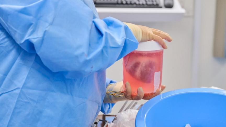
Takeaways * Scientists have made progress growing human liver in the lab. * The challenge has been to direct stems cells to grow into a mature, functioning adult organ. * This study shows that stem cells can be programmed, using genetic engineering, to grow from immature cells into mature tissue. * When a tiny lab-grown liver was transplanted into mice with liver disease, it extended the lives of the sick animals.* * *Imagine if researchers could program stem cells, which have the potential to grow into all cell types in the body, so that they could generate an entire human organ. This would allow scientists to manufacture tissues for testing drugs and reduce the demand for transplant organs by having new ones grown directly from a patient’s cells. I’m a researcher working in this new field – called synthetic biology – focused on creating new biological parts and redesigning existing biological systems. In a new paper, my colleagues and I showed progress in one of the key challenges with lab-grown organs – figuring out the genes necessary to produce the variety of mature cells needed to construct a functioning liver. Induced pluripotent stem cells, a subgroup of stem cells, are capable of producing cells that can build entire organs in the human body. But they can do this job only if they receive the right quantity of growth signals at the right time from their environment. If this happens, they eventually give rise to different cell types that can assemble and mature in the form of human organs and tissues. The tissues researchers generate from pluripotent stem cells can provide a unique source for personalized medicine from transplantation to novel drug discovery. But unfortunately, synthetic tissues from stem cells are not always suitable for transplant or drug testing because they contain unwanted cells from other tissues, or lack the tissue maturity and a complete network of blood vessels necessary for bringing oxygen and nutrients needed to nurture an organ. That is why having a framework to assess whether these lab-grown cells and tissues are doing their job, and how to make them more like human organs, is critical. Inspired by this challenge, I was determined to establish a synthetic biology method to read and write, or program, tissue development. I am trying to do this using the genetic language of stem cells, similar to what is used by nature to form human organs. Tissues and organs made by genetic designsI am a researcher specializing in synthetic biology and biological engineering at the Pittsburgh Liver Research Center and McGowan Institute for Regenerative Medicine, where the goals are to use engineering approaches to analyze and build novel biological systems and solve human health problems. My lab combines synthetic biology and regenerative medicine in a new field that strives to replace, regrow or repair diseased organs or tissues. I chose to focus on growing new human livers because this organ is vital for controlling most levels of chemicals – like proteins or sugar – in the blood. The liver also breaks down harmful chemicals and metabolizes many drugs in our body. But the liver tissue is also vulnerable and can be damaged and destroyed by many diseases, such as hepatitis or fatty liver disease. There is a shortage of donor organs, which limits liver transplantation. To make synthetic organs and tissues, scientists need to be able to control stem cells so that they can form into different types of cells, such as liver cells and blood vessel cells. The goal is to mature these stem cells into miniorgans, or organoids, containing blood vessels and the correct adult cell types that would be found in a natural organ. One way to orchestrate maturation of synthetic tissues is to determine the list of genes needed to induce a group of stem cells to grow, mature and evolve into a complete and functioning organ. To derive this list I worked with Patrick Cahan and Samira Kiani to first use computational analysis to identify genes involved in transforming a group of stem cells into a mature functioning liver. Then our team led by two of my students – Jeremy Velazquez and Ryan LeGraw – used genetic engineering to alter specific genes we had identified and used them to help build and mature human liver tissues from stem cells. The tissue is grown from a layer of genetically engineered stem cells in a petri dish. The function of genetic programs together with nutrients is to orchestrate formation of liver organoids over the course of 15 to 17 days. Liver in a dishI and my colleagues first compared the active genes in fetal liver organoids we had grown in the lab with those in adult human livers using a computational analysis to get a list of genes needed for driving fetal liver organoids to mature into adult organs. We then used genetic engineering to tweak genes – and the resulting proteins – that the stem cells needed to mature further toward an adult liver. In the course of about 17 days we generated tiny – several millimeters in width – but more mature liver tissues with a range of cells typically found in livers in the third trimester of human pregnancies. Like a mature human liver, these synthetic livers were able to store, synthesize and metabolize nutrients. Though our lab-grown livers were small, we are hopeful that we can scale them up in the future. While they share many similar features with adult livers, they aren’t perfect and our team still has work to do. For example, we still need to improve the capacity of the liver tissue to metabolize a variety of drugs. We also need to make it safer and more efficacious for eventual application in humans.[Deep knowledge, daily. Sign up for The Conversation’s newsletter.]Our study demonstrates the ability of these lab livers to mature and develop a functional network of blood vessels in just two and a half weeks. We believe this approach can pave the path for the manufacture of other organs with vasculature via genetic programming. The liver organoids provide several key features of an adult human liver such as production of key blood proteins and regulation of bile – a chemical important for digestion of food. When we implanted the lab-grown liver tissues into mice suffering from liver disease, it increased the life span. We named our organoids “designer organoids,” as they are generated via a genetic design. This article is republished from The Conversation, a nonprofit news site dedicated to sharing ideas from academic experts. It was written by: Mo Ebrahimkhani, University of Pittsburgh. Read more: * Brain organoids help neuroscientists understand brain development, but aren’t perfect matches for real brains * Why are scientists trying to manufacture organs in space?Mo Ebrahimkhani receives funding from National Institute of Health, University of Pittsburgh and Arizona Biomedical Research Council.

Finding alternatives to antibiotics is one of the biggest challenges facing the research community. Bacteria are increasingly resistant to these drugs, and this resistance leads to the deaths of more than 25,000 around the world. Now, a multidisciplinary team of researchers from the Universitat Rovira i Virgili, the University of Grenoble (France), the University of Saarland (Germany) and RMIT University (Australia) have discovered that the mechanical deformation of bacteria is a toxic mechanism that can kill bacteria with gold nanoparticles. The results of this research have been published in the journal Advanced Materials and are a breakthrough in researchers’ understanding the antibacterial effects of nanoparticles and their efforts to find new materials with bactericide properties.

Since the times of Ancient Egypt, gold has been used in a range of medical applications and, more recently, as for diagnosing and treating diseases such as cancer. This is due to the fact that gold is a chemically inert material, that is, it does not react or change when it comes into contact with an organism. Amongst the scientific community, nanoparticles are known for their ability to make tumors visible and for their applications in nanomedicine.

This new research shows that these chemically inert nanoparticles can kill thanks to a physical mechanism that deforms the cell wall. To demonstrate this, the researchers have synthesized in the laboratory in the shape of an almost perfect sphere and others in the shape of stars, all measuring 100 nanometres (8 times thinner than a hair). The group analyzed how these particle interact with living bacteria. “We find that the bacteria become deformed and deflate like a ball that is having the air let out before dying in the presence of these nanoparticles,” explained Vladimir Baulin, researcher at the Department of Chemical Engineering of the URV. The researchers state the bacteria seem to have died after a massive leak, “as if the cell wall had spontaneously exploded.”

Dr yu shrike zhang phd is assistant professor at harvard medical school and associate bioengineer at brigham and women’s hospital.

Dr. Zhang’s research interests include symbiotic tissue engineering, 3D bio-printing, organ-on-a-chip technology, biomaterials, regenerative engineering, bioanalysis, nanomedicine, and biology.

His scientific contributions have been recognized by over 40 regional, national and international awards. He has been invited to deliver more than 110 lectures worldwide, and has served as reviewer for more than 500 manuscripts for as many as 50 journals.

Dr. Zhang is serving as Editor-in-Chief for Microphysiological Systems, and is Associate Editor for Bio-Design and Manufacturing, Nano Select, Aggregate, and Essays in Biochemistry.

As the COVID-19 pandemic continues to spread across the world, testing remains a key strategy for tracking and containing the virus. Bioengineering graduate student, Maha Alafeef, has co-developed a rapid, ultrasensitive test using a paper-based electrochemical sensor that can detect the presence of the virus in less than five minutes. The team led by professor Dipanjan Pan reported their findings in ACS Nano.

“Currently, we are experiencing a once-in-a-century life-changing event,” said Alafeef. “We are responding to this global need from a holistic approach by developing multidisciplinary tools for early detection and diagnosis and treatment for SARS-CoV-2.”

There are two broad categories of COVID-19 tests on the market. The first category uses reverse transcriptase real-time polymerase chain reaction (RT-PCR) and nucleic acid hybridization strategies to identify viral RNA. Current FDA-approved diagnostic tests use this technique. Some drawbacks include the amount of time it takes to complete the test, the need for specialized personnel and the availability of equipment and reagents.

Ineurals — advanced neuro-technologies for rapid learning and skill acquisition.


The 711th Human Performance Wing, under the U.S. Air Force Research Laboratory leads the development, integration, and delivery of Airman-centric research, education, and consultation enabling the U.S. Air Force to achieve responsive and effective global vigilance, global reach, and global power now and in the future. It’s comprised of the United States Air Force School of Aerospace Medicine and the Airman Systems Directorate, whose science and technology competencies include Training, Adaptive Warfighter Interfaces, Bioeffects, Bioengineering, and Aerospace and Operational Medicine.

The Individualized Neural Learning System, or iNeuraLS, is a new augmented learning platform that will enable rapid learning by closed-loop modulation of cognitive states during skill acquisition. Essentially, the AFRL team seeks to develop a capability that will give Airmen the ability to rapidly acquire knowledge and skills on the fly through direct brain interfaces with the help of neurotechnologies.

Although Drosophila is an insect whose genome has only about 14,000 genes, roughly half the human count, a remarkable number of these have very close counterparts in humans; some even occur in the same order in the fly’s DNA as in our own. This, plus the organism’s more than 100-year history in the lab, makes it one of the most important models for studying basic biology and disease.

To take full advantage of the opportunities offered by Drosophila, researchers need improved tools to manipulate the fly’s genes with precision, allowing them to introduce mutations to break genes, control their activity, label their protein products, or introduce other inherited genetic changes.

“We now have the genome sequences of lots of different animals — worms, flies, fish, mice, chimps, humans,” says Roger Hoskins of Berkeley Lab’s Life Sciences Division. “Now we want improved technologies for introducing precise changes into the genomes of lab animals; we want efficient genome engineering. Methods for doing this are very advanced in bacteria and yeast. Good methods for worms, flies, and mice have also been around for a long time, and improvements have come along fairly regularly. But with whole genome sequences in hand, the goals are becoming more ambitious.”

O,.o Circa 2019


CRISPR/Cas9 is now a household name associated with genetic engineering studies. Through cutting-edge research described in their paper published in Scientific Reports, a team of researchers from Tokyo University of Science, Meiji University, and Tokyo University of Agriculture and Technology, led by Dr Takayuki Arazoe and Prof Shigeru Kuwata, has recently established a series of novel strategies to increase the efficiency of targeted gene disruption and new gene “introduction” using the CRISPR/Cas9 system in the rice blast fungus Pyricularia (Magnaporthe) oryzae. These strategies include quicker (single-step) gene introduction, use of small homologous sequences, and bypassing of certain prerequisite host DNA “patterns” and host component modification.

The team led by Dr Arazoe and Prof Kuwata has devised simple and quick techniques for gene editing (target gene disruption, sequence substitution, and re-introduction of desired genes) using CRISPR/Cas9 in the rice blast fungus Pyricularia (Magnaporthe) oryzae, a type of filamentous fungus. Spurred on by encouraging results, the researchers surmise, “Plants and their pathogens are still coevolving in nature. Exploiting the mutation mechanisms of model pathogenic fungi as a genome editing technique might lead to the development of further novel techniques in genetic engineering.”

The working component of the CRISPR/Cas9 system binds to the target gene region (DNA) and causes a site-specific double-stranded break (DSB) in the DNA. Effective binding of this component requires a certain “motif” or “pattern” called the protospacer-adjacent motif (PAM), which follows downstream of the target gene region.

The honeybee (Apis mellifera) is an important insect pollinator of wild flowers and crops, playing critical roles in the global ecosystem. Additionally, the honeybee serves as an ideal social insect model. Therefore, functional studies on honeybee genes are of great interest. However, until now, effective gene manipulation methods have not been available in honeybees. Here, we reported an improved CRISPR/Cas9 gene-editing method by microinjecting sgRNA and Cas9 protein into the region of zygote formation within 2 hr after queen oviposition, which allows one-step generation of biallelic knockout mutants in honeybee with high efficiency. We first targeted the Mrjp1 gene. Two batches of honeybee embryos were collected and injected with Mrjp1 sgRNA and Cas9 protein at the ventral cephalic side and the dorsal posterior side of the embryos, respectively. The gene-editing rate at the ventral cephalic side was 93.3%, which was much higher than that (11.8%) of the dorsal-posterior-side injection. To validate the high efficiency of our honeybee gene-editing system, we targeted another gene, Pax6, and injected Pax6 sgRNA and Cas9 protein at the ventral cephalic side in the third batch. A 100% editing rate was obtained. Sanger sequencing of the TA clones showed that 73.3% (for Mrjp1) and 76.9% (for Pax6) of the edited current-generation embryos were biallelic knockout mutants. These results suggest that the CRISPR/Cas9 method we established permits one-step biallelic knockout of target genes in honeybee embryos, thereby demonstrating an efficient application to functional studies of honeybee genes. It also provides a useful reference to gene editing in other insects with elongated eggs.