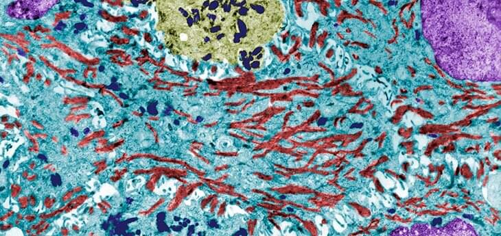
A new UC Davis-led study sheds light on cell type-specific biomarkers, or signs, of melanoma. The research was recently published in the Journal of Investigative Dermatology.
Melanoma, the deadliest of the common skin cancers, is curable with early diagnosis and treatment. However, diagnosing melanoma clinically and under the microscope can be complicated by what are called melanocytic nevi—otherwise known as birth marks or moles that are non-cancerous. The development of melanoma is a multi-step process where “melanocytes,” or the cells in the skin that contain melanin, mutate and proliferate. Properly identifying melanoma at an early stage is critical for improved survival.
“The biomarkers of early melanoma evolution and their origin within the tumor and its microenvironment are a potential key to early diagnosis of melanoma,” said corresponding author of the study Maija Kiuru, associate professor of clinical dermatology and pathology at UC Davis Health. “To unravel the mystery, we used high-plex spatial RNA profiling to capture distinct gene expression patterns across cell types during melanoma development. This approach allows studying the expression of hundreds or thousands of genes without disrupting the native architecture of the tumor.”
