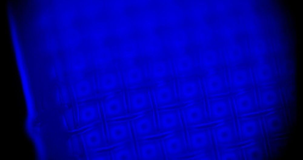
Curtin University researchers have discovered a new way to more accurately analyze microscopic samples by essentially making them glow in the dark through the use of chemically luminescent molecules.
Lead researcher Dr. Yan Vogel from the School of Molecular and Life Sciences said current methods of microscopic imaging rely on fluorescence, which means a light needs to be shining on the sample while it is being analyzed. While this method is effective, it also has some drawbacks.
“Most biological cells and chemicals generally do not like exposure to light because it can destroy things—similar to how certain plastics lose their colors after prolonged sun exposure, or how our skin can get sunburned,” Dr. Vogel said. “The light that shines on the samples is often too damaging for the living specimens and can be too invasive, interfering with the biochemical process and potentially limiting the study and scientists’ understanding of the living organisms.”
